. 2005 Nov 8;102(45):16327-32.
doi: 10.1073/pnas.0508335102. Epub 2005 Oct 31.

An immune-responsive serpin, SRPN6, mediates mosquito defense against malaria parasites

Affiliations

An immune-responsive serpin, SRPN6, mediates mosquito defense against malaria parasites

Eappen G Abraham et al. Proc Natl Acad Sci U S A. .

Abstract

We have functionally analyzed the orthologous SRPN6 genes from Anopheles stephensi and Anopheles gambiae using phylogenetic, molecular, reverse genetic, and cell biological tools. The results strongly implicate SRPN6 in the innate immune response against Plasmodium. This gene belongs to a mosquito-specific gene cluster including three additional Anopheles serpins. SRPN6 expression is induced by Escherichia coli and both rodent and human malaria parasites. The gene is specifically expressed in midgut cells invaded by Plasmodium ookinetes and in circulating and attached hemocytes. Knockdown of SRPN6 expression by RNA interference in susceptible An. stephensi leads to substantially increased parasite numbers, whereas depletion in susceptible An. gambiae delays progression of parasite lysis without affecting the number of developing parasites. However, the An. gambiae SRPN6 knockdown increases the number of melanized parasites in the L3-5 refractory strain and in susceptible G3 mosquitoes depleted of CTL4. These results indicate that AsSRPN6 is involved in the parasite-killing process, whereas AgSRPN6 acts on parasite clearance by inhibiting melanization and/or promoting parasite lysis. We propose that these observed phenotypic differences are due to changed roles of the respective target serine proteases in the two mosquito species.


Figures

Fig. 1.
Fig. 1.
Phylogenetic analysis of the SRPN6 mosquito-specific expansion cluster. (A) Bayesian inference of phylogenetic relationships of arthropod serpins related to SRPN6. The An. gambiae SRPN4, -5, -6, and -16 form a mosquito-specific expansion cluster together with An. stephensi SRPN6 and Ae. aegypti TC47107 and TC39371. Red labels correspond to mosquito, blue to drosophilid, and gray to honey bee sequences. ANGA, An. gambiae; ANST, An. stephensi; DRME, D. melanogaster; DRPS, Drosophila pseudoobscura; APME, Apis mellifera. Chromosomal arm (3R) locations are specified in Mb. Numbers on each branch indicate posterior probability (Upper) and bootstrap values (Lower). (B) Sequence alignment of the reactive center loops. The P1–P1′ residues immediately flank the arrow (scissile bond).
Fig. 2.
Fig. 2.
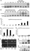
AsSRPN6 and AgSRPN6 are strongly up-regulated by immune challenge. (A) AsSRPN6 expression at different developmental stages and tissues and after noninfectious blood meal was not detected by Northern analysis. Positive control (+): Midguts 24 h after P. berghei infection. (B) Northern analysis revealed strong up-regulation of AsSRPN6 expression in midgut tissues 18–48 h after infection with gametocyte-forming P. berghei. (CE) qRT-PCR analysis of AgSRPN6 expression during development (C) and after P. berghei infection (D and E). Expression of AgSRPN6 was detected mainly in adults and was strongly up-regulated in midgut tissues 20–24 h postinfection (weakly so in 48-hpi carcasses). Data were normalized to S7 expression and calibrated to the average of all developmental stages or 18 h blood fed (BF). (F) RT-PCR analysis of AsSRPN6 induction by P. falciparum. PCR amplifications were performed by using 25 cycles for P. berghei and 30 cycles for P. falciparum experiments. Prevalence and parasite load for each experiment are indicated. (Lower) Amplification of the ribosomal protein S7 loading control. (G) Northern analysis of An. gambiae midgut tissue total RNA 6 h after ingestion of 3 × 10⁵ E. coli or ONN virus (H) ONN virus infection was verified by using a virus-specific probe (ONN). Ac, adult carcasses; BF, blood-fed; B, buffer-fed; E, embryos, Fnaive, sugar-fed females; L1–3, first to fourth instar larvae; L4g, fourth instar guts; L4c, fourth instar carcasses; ON, ONN-infected; ONc, ONN-infected carcass; U, uninfected; P, pupae; Pb, P. berghei-infected; Pf1 and Pf2, P. falciparum-infected (two independent experiments).
Fig. 3.
Fig. 3.
Immunolocalization and immunoblotting of As- and AgSRPN6 proteins. Localization of AsSRPN6 (A) and AgSRPN6 (B) by confocal microscopy in midgut sheets at indicated time points after P. berghei infection (Pb). Invading parasites were mostly in close proximity or within SRPN6-positive cells (A, Pb24h-i, Pb24h-ii); the number of AgSRPN6-positive cells dropped strongly by 48 hpi. Parasites were either visualized with monoclonal anti-P28 antibody (A) or by endogenous GFP expression (B). No SRPN6 protein was detected in midgut epithelia after noninfected blood feeding (BF). Arrows indicate invading parasites. Examples of SRPN6-positive midgut epithelial cells are marked by asterisks. In A, Pb24h-ii and B, Pb27h-1, a parasite is seen emerging from a previously invaded cell, which stains intensely for SRPN6 (asterisk). (Scale bars = 10μmin A Pb24h-ii and Pb27h-i; all other scale bars in A and B = 20μm.) (C) Immunoblot of An. stephensi midgut sheets before (U) and after either a noninfected (BF) or P. berghei-infected (Pb, 90% infection prevalence, mean oocysts number 200 per gut) blood meal. Numbers after BF and Pb indicate hours after blood ingestion. The equivalent of two gut sheets was loaded per lane. Rec, 25 ng of bacterially expressed thioredoxin-AgSRPN6 fusion protein. A ≈55-kDa protein band was detected only in infected midgut sheets. (D) AgSRPN6 protein was detected in immunoblots of conditioned medium of an An. gambiae cell line, indicating that the protein is secreted. Rec, 10 ng of bacterially expressed full-length AgSRPN6. (E) AgSRPN6 is expressed in circulating hemocytes. (Scale bar = 10 μm.) (F) 30° y-plane projection of a confocal stack reveals expression of AgSRPN6 in some small-nucleated cells attached to the hemolymph-facing basal side of the midgut epithelium. Filled arrowhead, nucleus of a midgut epithelial cell; open arrowhead, a small-nucleated cell. (Scale bar = 10 μm.) (G) Confocal slice (2 μm) of the same cell. (H) Constitutive presence of AgSRPN6 in pericardial cells. (Scale bar = 50 μm.)
Fig. 4.
Fig. 4.
Effects of SRPN6 KD on P. berghei development in the mosquito. (AD) Graphs show the distribution of overall numbers of parasites per midgut 8–15 days postinoculation in dsSRPN6 or control dsGFP-treated mosquitoes. Geometric means (±1 SE) in the pooled datasets from at least three independent experiments, the numbers of midguts examined, and the percentage of melanized parasites per midgut are indicated. See also Table 3 and Fig. 6, which is published as supporting information on the PNAS web site. The SRPN6-KD significantly increases the number of developing parasites in susceptible An. stephensi (A), but not in susceptible An. gambiae (B and D); the numbers of melanized parasites increase significantly in dsSRPN6 vs. control-treated refractory L3-5 (C) and in the double-KD CTL4/SRPN6 An. gambiae (D). (E) Three-dimensional projections of midguts dissected and stained with anti-P28 antibody and DAPI at indicated times after infection. Mosquitoes had been treated with either dsAgSRPN6 or dsGFP 4 days before infection. Living parasites (arrows) appear green and yellow, because they are double-labeled with anti-P28 antibody (red) and endogenous GFP (green). Open arrowheads indicate P28-only parasites in the process of lysis. White squares, parasites are shown in close-up insets in F. (Scale bar = 20 m.) (F) Graphical representation of temporally changing parasite phenotypes detected in dsGFP and dsSRPN6-treated mosquito midguts at indicated times after infection. Bar graphs, coded as indicated in the insets, represent GFP-fluorescent live parasites (green) or lysing P28-only parasites (red). (Insets) Confocal micrographs of these two parasite classes. Error bars represent standard error in the pooled data set of three independent experiments.

References

    1. Snow, R. W., Guerra, C. A., Noor, A. M., Myint, H. Y. & Hay, S. I. (2005) Nature 434, 214-217. - PMC - PubMed
    1. Niare, O., Markianos, K., Volz, J., Oduol, F., Toure, A., Bagayoko, M., Sangare, D., Traore, S. F., Wang, R., Blass, C., et al. (2002) Science 298, 213-216. - PubMed
    1. Sinden, R. E. (2002) Cell. Microbiol. 4, 713-724. - PubMed
    1. Collins, F. H., Sakai, R. K., Vernick, K. D., Paskewitz, S., Seeley, D. C., Miller, L. H., Collins, W. E., Campbell, C. C. & Gwadz, R. W. (1986) Science 234, 607-610. - PubMed
    1. Vernick, K. D., Fujioka, H., Seeley, D. C., Tandler, B., Aikawa, M. & Miller, L. H. (1995) Exp. Parasitol. 80, 583-595. - PubMed

Publication types

LinkOut - more resources