. 2005 Nov 8;102(45):16309-14.
doi: 10.1073/pnas.0508237102. Epub 2005 Oct 31.

IL-32 synergizes with nucleotide oligomerization domain (NOD) 1 and NOD2 ligands for IL-1beta and IL-6 production through a caspase 1-dependent mechanism

Affiliations

IL-32 synergizes with nucleotide oligomerization domain (NOD) 1 and NOD2 ligands for IL-1beta and IL-6 production through a caspase 1-dependent mechanism

Mihai G Netea et al. Proc Natl Acad Sci U S A. .

Abstract

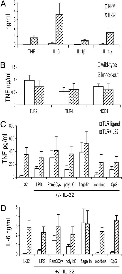
The activation of innate immunity requires the amplification of signals induced by pattern-recognition receptors for bacterial products. We have investigated the role of the newly described cytokine IL-32 in the amplification of cytokine production induced by the two most clinically relevant families of microbial receptors, the cell-surface Toll-like receptors (TLRs) and the intracellular nuclear oligomerization domain (NOD) receptor family. IL-32 synergized with the NOD1- and NOD2-specific muropeptides of peptidoglycans for the release of IL-1beta and IL-6 (a 3- to 10-fold increase). In contrast, IL-32 did not influence the cytokine production induced via TLRs. The synergistic effect of IL-32 and synthetic muramyl dipeptide (MDP) on cytokine production was absent in the cells of patients with Crohn's disease bearing the NOD2 frameshift mutation 3020insC, demonstrating that the IL-32/MDP synergism depends on NOD2. This in vitro synergism between IL-32 and NOD2 ligands was consistent with a marked constitutive expression of IL-32 in human colon epithelial tissue. In addition, the potentiating effect of IL-32 on the cytokine production induced by the synthetic muropeptide FK-156 was absent in NOD1-deficient macrophages, supporting the interaction between IL-32 and NOD1 pathways. When specific caspase inhibitors were used, the synergism between IL-32 and MDP/NOD2 depended on the activation of caspase 1. Only additive effects of IL-32 and muropeptides were observed for TNF-alpha production. The modulation of intracellular NOD2 pathways by IL-32, but not cell-surface TLRs, and the marked expression of IL-32 in colon mucosa suggest a role of IL-32 in the pathogenesis of Crohn's disease.


Figures

Fig. 1.
Fig. 1.
IL-32 interaction with TLR pathways. (A) PBMCs were stimulated with IL-32γ (20 ng/ml) (hatched bars), and after 24 h the concentrations of TNF-α, IL-6, IL-1β, and intracellular IL-1α were measured. The presence of RPMI medium 1640 indicates unstimulated cells. (B) Peritoneal macrophages from TLR2-/-, TLR4-/-, and NOD1-/- mice (hatched bars) and the respective controls (open bars) were stimulated with 20 ng/ml IL-32γ.(C and D) Ligands of TLR4 (LPS), TLR2 (Pam3Cys), TLR3 [(poly(I)·poly(C)], TLR5 (flagellin), TLR7 (loxoribine), and TLR9 (CpG DNA) were added to PBMCs (open bars), with or without costimulation with IL-32 (hatched bars). Data are presented as means ± SEM (n = 7).
Fig. 2.
Fig. 2.
IL-32 amplification of cytokine production stimulated by muropeptides. (AD) In human PBMCs, IL-32γ (10 ng/ml) (hatched bars) was coincubated with MDP or Mur-Tri-DAP (both at 1μg/ml), and production of IL-1β (A), IL-6 (B), TNF-α (C), and IL-1α (D) is shown. MDP or Mur-Tri-DAP stimulation alone is represented by open bars. (E). The effects on IL-6 production when using different concentrations of IL-32 and MDP are shown. Open bars represent PBMCs stimulated with 5ng/ml IL-32γ plus the corresponding MDP concentration inμg/ml; hatched bars represent PBMCs stimulated with 10 ng/ml IL-32γ plus the corresponding MDP concentration; solid bars represent 20 ng/ml IL-32γ plus MDP; stippled bars represent 50 ng/ml IL-32γ plus MDP concentration. The data are presented as fold increase in which the nanograms of IL-32-induced IL-6 and the nanograms of MDP-induced IL-6 from individual PBMC cultures were added and assigned a value of 1.0. The measured value of IL-6 induced by the combination of IL-32γ plus MDP was then calculated as fold increase over 1.0. Data are presented as means ± SEM (n = 7). *, P < 0.01.
Fig. 3.
Fig. 3.
Monocyte-dependent IL-32/MDP synergism. PBMCs, adherent monocytes, or nonadherent lymphocytes were stimulated with MDP/IL-32 as described for Fig. 2. After 24 h, IL-1β (A) and IL-6 (B) were measured. Data are presented as means ± SD (n = 7). *, P < 0.01.
Fig. 4.
Fig. 4.
The synergism among IL-32 and muropeptides is mediated by NOD1 and NOD2. (A) PBMCs isolated from five healthy controls, five Crohn's patients bearing the WT NOD2 allele (Crohn-WT), and four patients homozygous for the frameshift mutation 3020insC NOD2 (Crohn-FS) were stimulated with combinations of IL-32γ (10 ng/ml) and MDP (10 μg/ml) for 24 h. Data are presented as means ± SEM. (B) IL-32γ (10 ng/ml) and the murine NOD1 ligand FK-156 (1 μM) were added together to peritoneal macrophages harvested from control NOD1+/+ mice and NOD1-/- animals. After 24 h, IL-6 concentration was measured. Data are presented as means ± SEM (n = 10). *, P < 0.05.
Fig. 5.
Fig. 5.
The role of proinflammatory caspases for the IL-32 effect on NOD2 stimulation. A pan-caspase inhibitor, a caspase 1 inhibitor, or a caspase 1 and 5 inhibitor (each at 20 μM) was added to the stimulation of PBMCs with IL-32γ (open bars), MDP (hatched bars), or a combination of both (filled bars). (AC) Concentrations of IL-1β (A), IL-6 (B), and TNF-α (C) were measured after a 24-h stimulation. (D) IL-1Ra (10 μg/ml) or IL-18BP (10 μg/ml) was added to the IL-32/MDP stimulation. Data are presented as means ± SEM (n = 7). *, P < 0.05.
Fig. 6.
Fig. 6.
IL-32 expression in the epithelial cells of colon mucosa. Biopsies of the colon mucosa of human large intestine were stained with a murine anti-human IL-32 monoclonal antibody. The staining of colon tissue of one representative individual of four healthy volunteers tested is shown. IL-32 is largely expressed in the differentiated colon epithelial cells, whereas the control antibody showed no staining. (Magnification: A, ×200; B, ×400. Scale bar: 100 μm.)

References

    1. Kim, S. H., Han, S. Y., Azam, T., Yoon, D. Y. & Dinarello, C. A. (2005) Immunity 22, 131-142. - PubMed
    1. Medzhitov, R. (2001) Nat. Rev. Immunol. 1, 135-145. - PubMed
    1. Beutler, B. (2004) Nature 430, 257-263. - PubMed
    1. Kapsenberg, M. (2003) Nat. Rev. Immunol. 3, 984-993. - PubMed
    1. Inohara, N., Ogura, Y. & Nunez, G. (2002) Curr. Opin. Microbiol. 5, 76-80. - PubMed

Publication types

MeSH terms