Comparative Study
. 2005 Nov 29;102(48):17308-13.
doi: 10.1073/pnas.0506924102. Epub 2005 Oct 31.

Structural basis for the recognition between HIV-1 integrase and transcriptional coactivator p75

Affiliations
Comparative Study

Structural basis for the recognition between HIV-1 integrase and transcriptional coactivator p75

Peter Cherepanov et al. Proc Natl Acad Sci U S A. .

Abstract

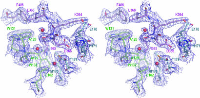
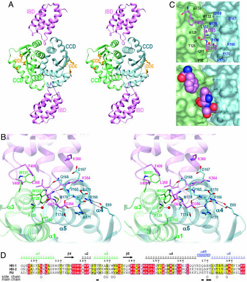
Integrase (IN) is an essential retroviral enzyme, and human transcriptional coactivator p75, which is also referred to as lens epithelium-derived growth factor (LEDGF), is the dominant cellular binding partner of HIV-1 IN. Here, we report the crystal structure of the dimeric catalytic core domain of HIV-1 IN complexed to the IN-binding domain of LEDGF. Previously identified LEDGF hotspot residues anchor the protein to both monomers at the IN dimer interface. The principal structural features of IN that are recognized by the host factor are the backbone conformation of residues 168-171 from one monomer and a hydrophobic patch that is primarily comprised of alpha-helices 1 and 3 of the second IN monomer. Inspection of diverse retroviral primary and secondary sequence elements helps to explain the apparent lentiviral tropism of the LEDGF-IN interaction. Because the lethal phenotypes of HIV-1 mutant viruses unable to interact with LEDGF indicate that IN function is highly sensitive to perturbations of the structure around the LEDGF-binding site, we propose that small molecule inhibitors of the protein-protein interaction might similarly disrupt HIV-1 replication.


Figures

Fig. 1.
Fig. 1.
Electron density map for residues at the CCD–IBD interface. Selected interface residues are shown as sticks. The final 2FoFc electron density map (dark blue) is at a 1-σ contour level. The color code for carbon atoms is: violet, LEDGF chain C; blue, IN chain A; green, IN chain B. Three water molecules buried at the protein interface are shown as red spheres.
Fig. 2.
Fig. 2.
Molecular mechanism of the IN–LEDGF interaction. (A) The overall structure of the CCD–IBD complex. IN chains A and B are colored blue and green, respectively; the IBD subunits are violet. The side chains of the DDE catalytic triad are shown as yellow sticks. (B) Key intermolecular contacts at the CCD–IBD interface. Selected residues are shown as sticks. The water molecule hydrogen-bonded to main-chain carbonyl groups of LEDGF residue Ile-365 and IN residue Thr-125 is shown as a red sphere. Hydrogen bonds discussed in the text and the salt bridge between IN residues Glu-69 and Arg-166 are indicated by dotted lines. (C) The pocket at the CCD dimer interface. LEDGF hotspot residues Ile-365, Asp-366, and Phe-406 are shown as sticks (Upper) or in space-fill mode (Lower). The IN subunits are shown as semitransparent surfaces. Selected IN residues are indicated. (D) Sequence alignment of HIV-1, HIV-2, and feline immunodeficiency virus (FIV) INs. Identical residues are white on red background; residues with conserved properties are bold on yellow background. Residue numbering, secondary structure elements, and the position of the α4/5 connector in HIV-1 IN are shown above the alignment; structural elements are colorized as in AC. Open circles and filled boxes under the alignment indicate residues that make contacts to the LEDGF IBD through side-chain and main-chain atoms, respectively. The alignment was printed by using espript-2.2 (56).
Fig. 3.
Fig. 3.
Molecular details at the dimer interfaces of HIV-1 and RSV IN. (A) Comparison of the IN CCD in complex with LEDGF to the unliganded CCD structure. The structure of the CCD–IBD complex was superimposed onto the structure of the free CCD from ref. (PDB code 1BL3). The color code for IN chains A and B is the same as in Figs. 1 and 2; the unliganded structure is painted brown. Selected side chains are shown as sticks. (B) Structure of the corresponding region of RSV IN (PDB code 1C0M, ref. 48). Chain A is painted green; chain B is blue; and the loop in chain B connecting α4 and α5 is shown in pink. Side chains of exposed hydrophobic and aromatic residues at the interface are shown as sticks.

Comment in

References

    1. Craigie, R. (2002) in Mobile DNA II, eds. Craig, N. L., Craigie, R., Gellert, M. & Lambowitz, A. M. (Am. Soc. Microbiol., Washington, DC), pp. 613–630.
    1. Turlure, F., Devroe, E., Silver, P. A. & Engelman, A. (2004) Front. Biosci. 9, 3187–3208. - PubMed
    1. Engelman, A. (2005) Proc. Natl. Acad. Sci. USA 102, 1275–1276. - PMC - PubMed
    1. Bushman, F. D. (2003) Cell 115, 135–138. - PubMed
    1. Engelman, A. & Craigie, R. (1992) J. Virol. 66, 6361–6369. - PMC - PubMed

Publication types

MeSH terms

Associated data