. 2005 Dec;14(12):2993-3003.
doi: 10.1110/ps.051622005. Epub 2005 Oct 31.

Disruption of an intermonomer salt bridge in the p53 tetramerization domain results in an increased propensity to form amyloid fibrils

Affiliations

Disruption of an intermonomer salt bridge in the p53 tetramerization domain results in an increased propensity to form amyloid fibrils

Charles Galea et al. Protein Sci. 2005 Dec.

Abstract

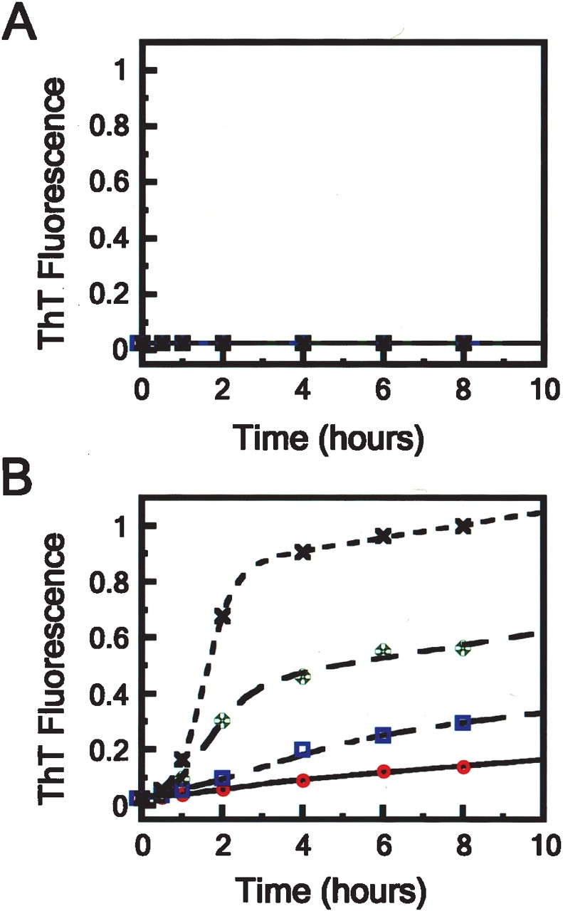
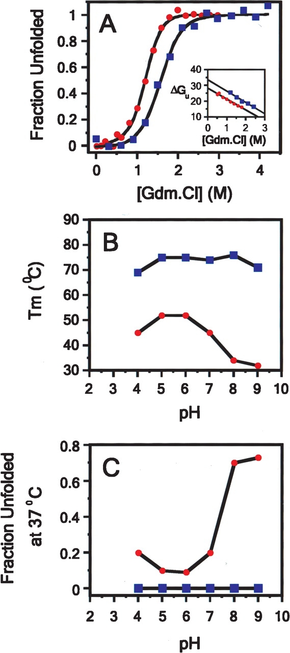
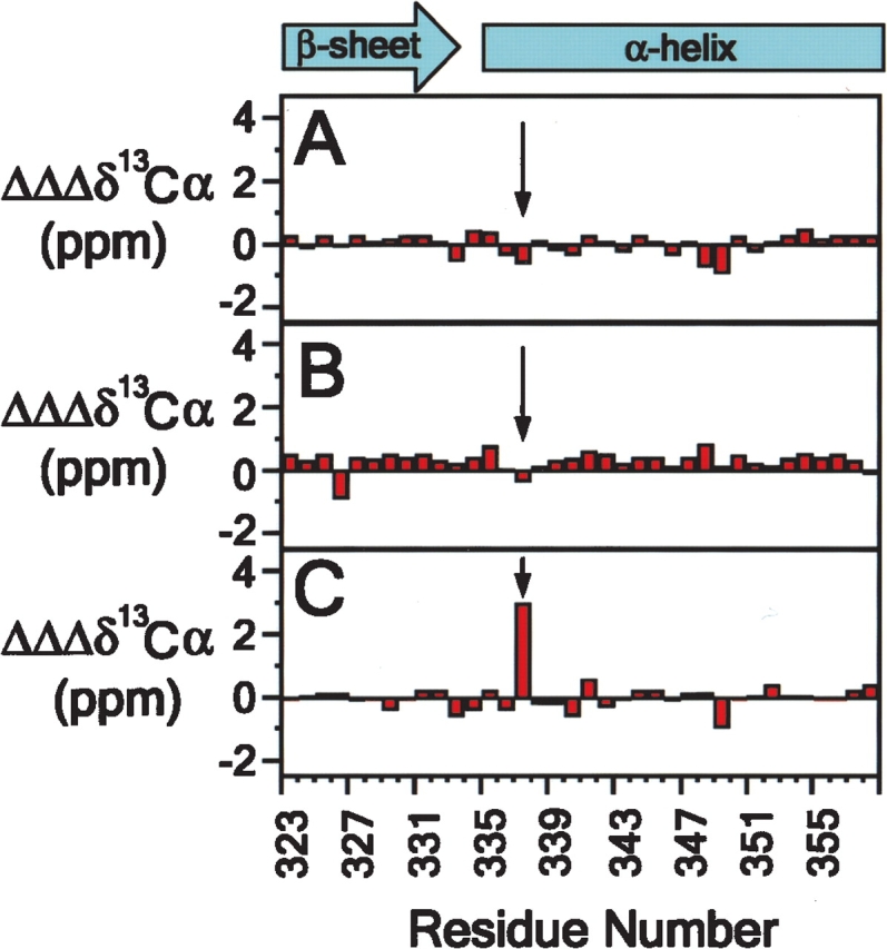
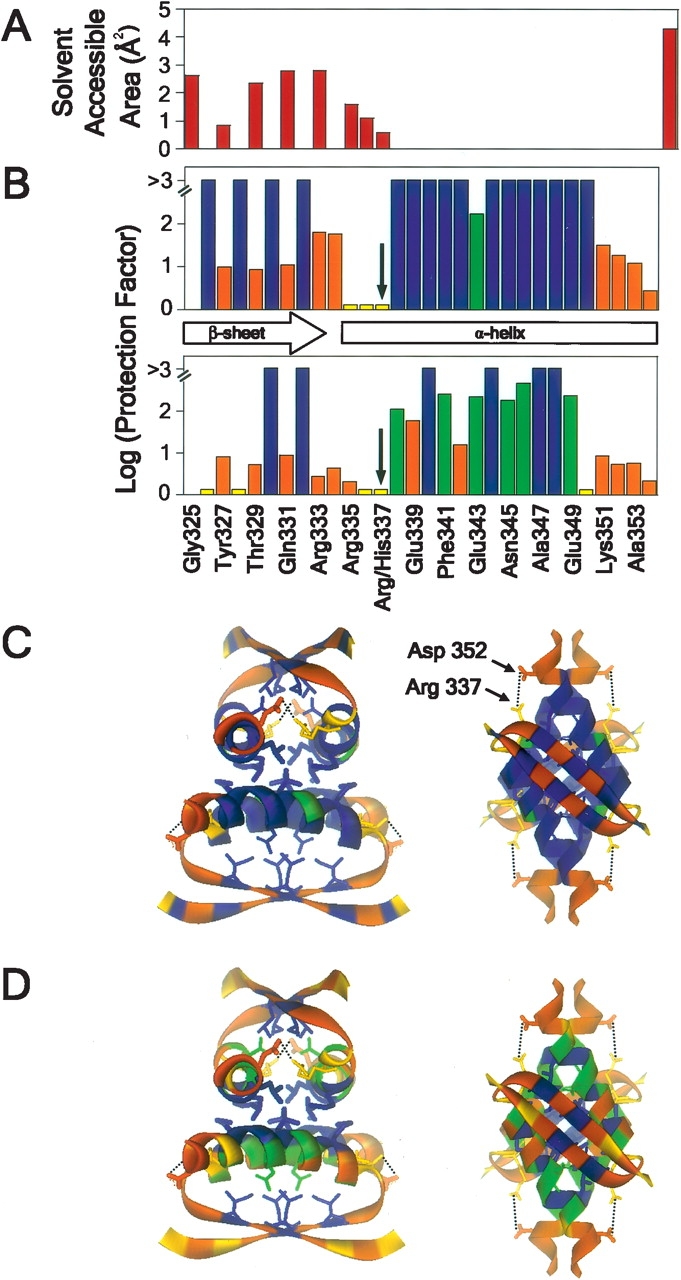
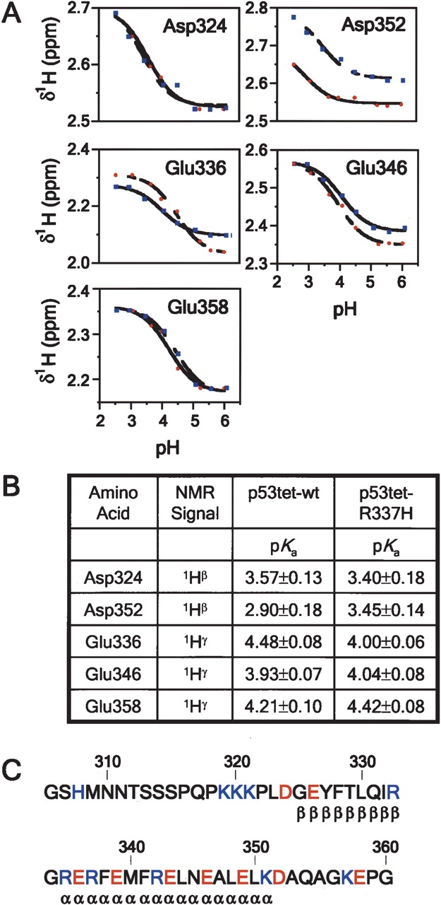
We describe in molecular detail how disruption of an intermonomer salt bridge (Arg337-Asp352) leads to partial destabilization of the p53 tetramerization domain and a dramatically increased propensity to form amyloid fibrils. At pH 4.0 and 37 degrees C, a p53 tetramerization domain mutant (p53tet-R337H), associated with adrenocortical carcinoma in children, readily formed amyloid fibrils, while the wild-type (p53tet-wt) did not. We characterized these proteins by equilibrium denaturation, 13C(alpha) secondary chemical shifts, (1H)-15N heteronuclear NOEs, and H/D exchange. Although p53tet-R337H was thermodynamically less stable, NMR data indicated that the two proteins had similar secondary structure and molecular dynamics. NMR derived pK(a) values indicated that at low pH the R337H mutation partially disrupted an intermonomer salt bridge. Backbone H/D exchange results showed that for at least a small population of p53tet-R337H molecules disruption of this salt bridge resulted in partial destabilization of the protein. It is proposed that this decrease in p53tet-R337H stability resulted in an increased propensity to form amyloid fibrils.


Figures

Figure 1.
Figure 1.
Influence of protein concentration upon the formation of amyloid fibrils by (A) p53tet-wt and (B) p53tet-R337H monitored by ThT fluorescence at pH 4.0 and 37°C. Protein concentrations were 1.0 (×), 0.5 (✠), 0.25 (□), and 0.13 (°) mM. Data were fitted to sigmoidal curves according to equation 8.
Figure 2.
Figure 2.
Guanidium and thermal denaturation studies of p53tet-wt (□) and p53tet-R337H (•) in 20 mM sodium phosphate buffer, pH 4.0, containing 50 mM NaCl at 25°C. (A) Guanidium denaturation curves for p53tet-wt and p53tet-R337H. The fraction of unfolded protein was determined using equation 2, and data were fitted according to equation 3. (Inset) Free energy of unfolding (ΔGu) in units of kcal mol−1 calculated using equation 4 plotted against denaturant concentration. (B) Variation of melting temperature (Tm) and (C) fraction unfolded protein (37°C) with pH for p53tet-wt and p53tet-R337H (DiGiammarino et al. 2002; Lee et al. 2003).
Figure 3.
Figure 3.
p53tet-wt and p53tet-R337H possess similar secondary structure over a range of pH values. Differences in secondary 13Cα chemical shift values for p53tet-wt (A) and p53tet-R337H (B) at pH 4.0 and 6.0 (pH 4.0 data minus pH 6.0 data) and secondary 13Cα chemical shift differences between p53tet-wt and p53tet-R337H (C) at pH 4.0 (p53tet-wt data minus p53tet-R337H data).
Figure 4.
Figure 4.
Difference in extent of backbone amide hydrogen exchange for p53tet-wt and p53tet-R337H at pH 4.0 and 20°C. (A) Surface-accessible area for backbone amide nitrogen atoms determined using the program GETAREA 1.1 (Fraczkiewicz and Braun 1998). (B) Log protection factor for residues in p53tet-wt (top) and p53tet-R337H (bottom). Ribbon diagram representation of the NMR solution structure of p53tet-wt (1PES; Lee et al. 1994) was used to illustrate the extent of hydrogen exchange for residues of (C) p53tet-wt and (D) p53tet-R337H using the program WebLab Viewer. Log protection factors >3 are shown in blue, between 0.1 and 3 are in green, and fast exchangers are in orange.
Figure 5.
Figure 5.
pKa values for acidic residues of p53tet-wt and p53tet-R337H. (A) Proton chemical shift titration curves for aspartate (1Hβ) and glutamate (1Hγ) residues of p53tet-wt (blue squares) and p53tet-R337H (red circles). Data were fit to the Henderson-Hasselbach equation as outlined under Materials and Methods. (B) Measured pKa values for acidic residues of p53tet-wt and p53tet-R337H. (C) Amino acid sequence of the wild-type p53 tetramerization domain. Acidic residues are shown in red and basic residues are shown in blue.

References

    1. Armstrong, K.M. and Baldwin, R.L. 1993. Charged histidine affects alpha-helix stability at all positions in the helix by interacting with the backbone charges. Proc. Natl. Acad. Sci. 90 11337–11340. - PMC - PubMed
    1. Bai, Y., Milne, J.S., Mayne, L., and Englander, S.W. 1993. Primary structure effects on peptide group hydrogen exchange. Proteins 17 75–86. - PMC - PubMed
    1. Bai, Y., Sosnick, T.R., Mayne, L., and Englander, S.W. 1995. Protein folding intermediates: Native-state hydrogen exchange. Science 269 192–197. - PMC - PubMed
    1. Booth, D.R., Sunde, M., Bellotti, V., Robinson, C.V., Hutchinson, W.L., Fraser, P.E., Hawkins, P.N., Dobson, C.M., Radford, S.E., Blake, C.C., et al. 1997. Instability, unfolding and aggregation of human lysozyme variants underlying amyloid fibrillogenesis. Nature 385 787–793. - PubMed
    1. Bosshard, H.R., Marti, D.N., and Jelesarov, I. 2004. Protein stabilization by salt bridges: Concepts, experimental approaches and clarification of some misunderstandings. J. Mol. Recognit. 17 1–16. - PubMed

Publication types