

Sema3D, Sema3F, and Sema5A are expressed in overlapping and distinct patterns in chick embryonic heart
- PMID: 16261621
- PMCID: PMC1768559
- DOI: 10.1002/dvdy.20614
Sema3D, Sema3F, and Sema5A are expressed in overlapping and distinct patterns in chick embryonic heart
Abstract
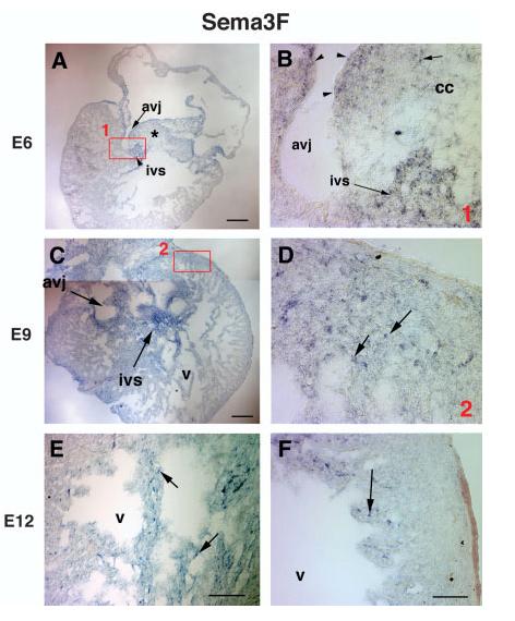
An increasing number of axon guidance cues have been shown recently to play important roles in the development of non-neural tissues. Semaphorins comprise one of the largest conserved families of axon guidance factors. We analyzed the expression patterns of Sema3D, Sema3F, and Sema5A genes in the chick embryonic heart by in situ hybridization. All three genes are expressed in the cardiac cushion regions, both in the mesenchymal cells, and epithelial cells in the endocardial layer, during the period of cardiac remodeling. In addition to the overlapping expression patterns in the cardiac cushion regions, these genes also exhibit distinct expression patterns in the developing heart: Sema3D is additionally expressed in the tips of the ventricular trabeculae; Sema3F is expressed in a subset of cells scattered throughout the ventricles; and Sema5A is expressed in the newly formed atrioventricular valves. The overlapping and distinct expression patterns of these genes suggest that they may play important roles in heart development.
2005 Wiley-Liss, Inc.
Figures


Similar articles
-
Sema3D and Sema7A have distinct expression patterns in chick embryonic development.Dev Dyn. 2006 Aug;235(8):2282-9. doi: 10.1002/dvdy.20882. Dev Dyn. 2006. PMID: 16804892 Free PMC article.
-
Semaphorin5A expression in the developing chick telencephalon.Brain Res Bull. 2005 Sep 15;66(4-6):436-40. doi: 10.1016/j.brainresbull.2005.02.011. Epub 2005 Mar 11. Brain Res Bull. 2005. PMID: 16144627
-
Developmental expression of sema3G, a novel zebrafish semaphorin.Gene Expr Patterns. 2005 Jun;5(5):647-53. doi: 10.1016/j.modgep.2005.02.009. Epub 2005 Apr 11. Gene Expr Patterns. 2005. PMID: 15939377
-
Cranial expression of class 3 secreted semaphorins and their neuropilin receptors.Dev Dyn. 2003 Dec;228(4):726-33. doi: 10.1002/dvdy.10396. Dev Dyn. 2003. PMID: 14648849
-
Gene expression during cardiac development.Symp Soc Exp Biol. 1992;46:251-64. Symp Soc Exp Biol. 1992. PMID: 1364123 Review.
Cited by
-
Wnt9a Can Influence Cell Fates and Neural Connectivity across the Radial Axis of the Developing Cochlea.J Neurosci. 2017 Sep 13;37(37):8975-8988. doi: 10.1523/JNEUROSCI.1554-17.2017. Epub 2017 Aug 14. J Neurosci. 2017. PMID: 28821654 Free PMC article.
-
Intraretinal projection of retinal ganglion cell axons as a model system for studying axon navigation.Brain Res. 2008 Feb 4;1192:165-77. doi: 10.1016/j.brainres.2007.01.116. Epub 2007 Feb 2. Brain Res. 2008. PMID: 17320832 Free PMC article. Review.
-
Rare copy number variations in adults with tetralogy of Fallot implicate novel risk gene pathways.PLoS Genet. 2012;8(8):e1002843. doi: 10.1371/journal.pgen.1002843. Epub 2012 Aug 9. PLoS Genet. 2012. PMID: 22912587 Free PMC article.
-
Notch signaling plays a key role in cardiac cell differentiation.Mech Dev. 2006 Aug;123(8):626-40. doi: 10.1016/j.mod.2006.06.003. Epub 2006 Jul 14. Mech Dev. 2006. PMID: 16843648 Free PMC article.
-
Cohesin: an emerging master regulator at the heart of cardiac development.Mol Biol Cell. 2023 May 1;34(5):rs2. doi: 10.1091/mbc.E22-12-0557. Epub 2023 Mar 22. Mol Biol Cell. 2023. PMID: 36947206 Free PMC article.
References
-
- Behar O, Golden JA, Mashimo H, Schoen FJ, Fishman MC. Semaphorin III is needed for normal patterning and growth of nerves, bones and heart. Nature. 1996;383:525–528. - PubMed
-
- Ben-Shachar G, Arcilla RA, Lucas RV, Manasek FJ. Ventricular trabeculations in the chick embryo heart and their contribution to ventricular and muscular septal development. Circ Res. 1985;57:759–766. - PubMed
-
- Bruneau BG. Transcriptional regulation of vertebrate cardiac morphogenesis. Circ Res. 2002;90:509–519. - PubMed
Publication types
MeSH terms
Substances
Grants and funding
LinkOut - more resources
Full Text Sources
Molecular Biology Databases
