. 2005 Nov;113(11):1522-9.
doi: 10.1289/ehp.7983.

Some environmental contaminants influence motor and feeding behaviors in the ornate wrasse (Thalassoma pavo) via distinct cerebral histamine receptor subtypes

Affiliations

Some environmental contaminants influence motor and feeding behaviors in the ornate wrasse (Thalassoma pavo) via distinct cerebral histamine receptor subtypes

Giuseppina Giusi et al. Environ Health Perspect. 2005 Nov.

Erratum in

  • Environ Health Perspect. 2005 Dec;113(12):A807

Abstract

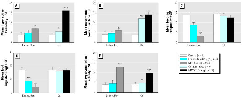
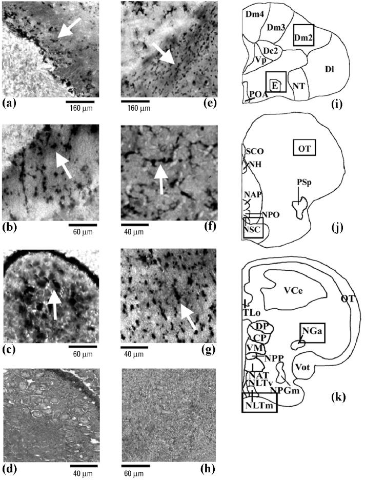
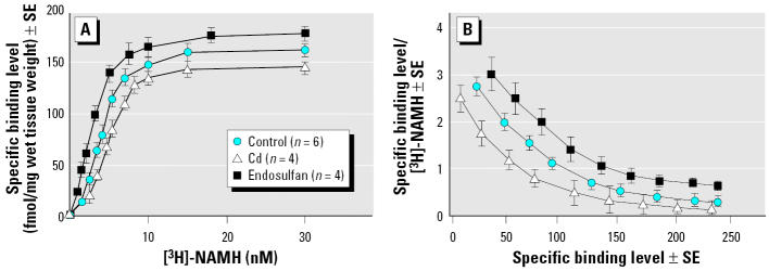
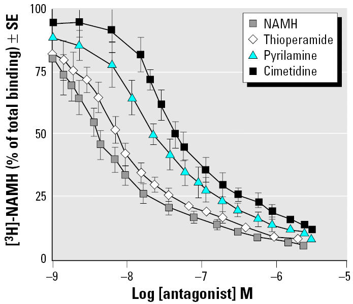
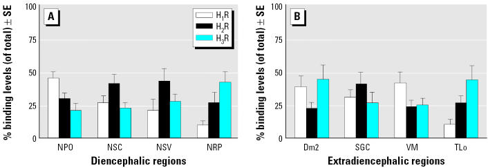
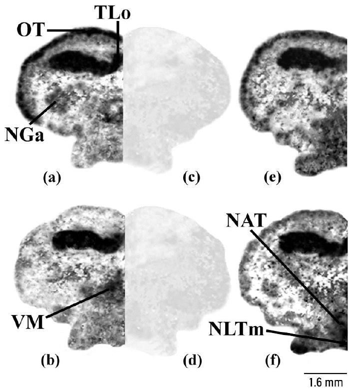
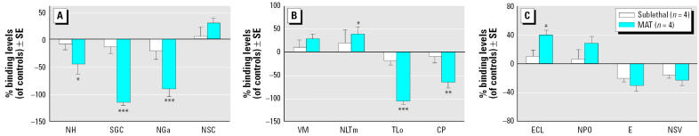
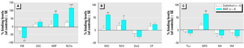
Common environmental contaminants such as heavy metals and pesticides pose serious risks to behavioral and neuroendocrine functions of many aquatic organisms. In the present study, we show that the heavy metal cadmium and the pesticide endosulfan produce such effects through an interaction of specific cerebral histamine receptor subtypes in the teleost ornate wrasse (Thalassoma pavo). Treatment of this teleost with toxic cadmium levels for 1 week was sufficient to induce abnormal swimming movements, whereas reduced feeding behaviors were provoked predominantly by elevated endosulfan concentrations. In the brain, these environmental contaminants caused neuronal degeneration in cerebral targets such as the mesencephalon and hypothalamus, damage that appeared to correlate with altered binding levels of the three major histamine receptors (subtypes 1, 2, and 3). Although cadmium accounted for reduced binding activity of all three subtypes in most brain regions, it was subtype 2 that seemed to be its main target, as shown by a very great (p < 0.001) down-regulation in mesencephalic areas such as the stratum griseum central layer. Conversely, endosulfan provided very great and great (p < 0.01) up-regulating effects of subtype 3 and 1 levels, respectively, in preoptic-hypothalamic areas such as the medial part of the lateral tuberal nucleus, and in the suprachiasmatic nucleus. These results suggest that the neurotoxicant-dependent abnormal motor and feeding behaviors may well be tightly linked to binding activities of distinct histamine subtypes in localized brain regions of the Thalassoma pavo.


Figures

Figure 1
Figure 1
Assessment of effects of Cd and endosulfan on some motor activities of Thalassoma pavo: (A) hyperactive movements, (B) movements toward surface, (C) feeding frequency, (D) quantity (mg) of food ingested, and (E ) hyperventilation activity. For these behaviors a sublethal concentration of endosulfan and Cd as well as a MAT concentration of the two contaminants, respectively, were compared with controls. Values (means of activities/24 hr ± SE) of motor activities and feeding behaviors were estimated daily during four 1-hr observations for 1 week, as described in “Materials and Methods.” The behavioral data were analyzed by one-way ANOVA followed where necessary by post hoc Neuman-Keuls multiple-range test. *p < 0.05; ***p < 0.001.
Figure 2
Figure 2
Photomicrographs showing the amino cupric silver staining pattern in rostral (i), middle (j), and posterior (k) brain areas of the Thalassoma pavo, treated with a MAT concentration of (a–c) Cd or (e–g) endosulfan. The effects of Cd (n = 6; arrows) were mostly observed in telencephalic and in mesencephalic areas such as Dm2 (a) and in the piriform SGC neurons of the optic tectum (b), respectively, and in the pretectal NGa (c), compared with control (n = 8); controls gave comparable results at all brain levels for both neurotoxicants as described in “Materials and Methods,” and so these same controls (d, h) were also used for the effects of endosulfan. In the case of endosulfan (n = 6), the major effects (arrows) were detected in the interneurons of the entopeduncular nucleus (e) and in the preoptic NSC area (f) and NLTm (g) of the hypothalamic lobe. Abbreviations: CP, central posterior thalamic nucleus; Dc2, central part of dorsal telencephalon, subdivision 2; Dl, lateral part of the dorsal telencephalon; Dm2–Dm4, medial part of the dorsal telencephalon, subdivisions 2–4; DP, dorsal posterior thalamic nucleus; E, entopeduncular nucleus; NAP, anterior periventricular nucleus; NAT, anterior tuberal nucleus; NGa, anterior part of the nucleus glomerulosus; NH, habenular nucleus; NLTm, medial part of lateral tuberal nucleus; NLTv, ventral part of lateral tuberal nucleus; NPGm, medial preglomerular nucleus; NPO, preoptic nucleus; NPP, posterior periventricular nucleus; NSC, suprachiasmatic nucleus; NT, nucleus taenia; OT, optic tectum; POA, preoptic area; PSp, parvocellular superficial pretectal nucleus; SCO, subcommissural organ; TLo, torus longitudinalis; VCe, cerebellum valvula; VM, ventromedial thalamic nucleus; Vot, ventral optic tract; Vp, postcommissural nucleus of the ventral telencephalon.
Figure 3
Figure 3
(A) A saturation curve of [3H]-NAMH binding (fmol/mg wet tissue weight ± SE), using wipe assays, was determined for the preoptic area of the Thalassoma pavo treated with MAT concentrations of Cd and endosulfan and compared with controls as described in “Materials and Methods.” (B) From the linear Scatchard plot, the negative slope was calculated to provide the mean dissociation constant (nM), whereas the intercept of the curve at the abscissa provided the maximal number of binding sites. Evaluation of saturation-binding study supplied similar results in three separate trials.
Figure 4
Figure 4
Displacement curves of [3H]-NAMH (% of total binding) in preoptic area of the Thalassoma pavo (n = 6) were generated in the presence of different concentrations (1 μM to 1 nm) of cold NAMH and of selective HA antagonists thioperamide, pyrilamine, and cimetidine as described in “Materials and Methods.” Each point represents mean ± SE of three separate tests.
Figure 5
Figure 5
Percentage binding levels (of total) ± SE of H1R, H2R, and H3R sites in diencephalic (A) and extra-diencephalic (B) regions of the Thalassoma pavo (n = 6) were determined in the presence of their respective selective antagonists as described in “Materials and Methods.” Abbreviations: Dm2, medial part of the dorsal telencephalon, subdivision 2; NPO, preoptic nucleus; NRP, nucleus of the posterior hypothalamic recess; NSC, suprachiasmatic nucleus; NSV, nucleus of the saccus vasculosus; SGC, stratum griseum central; TLo, torus longitudinalis; VM, ventromedial thalamic nucleus.
Figure 6
Figure 6
Representative binding autoradiograms displaying distinct receptor densities (black line) of H2R in the posterior regions of the Thalassoma pavo treated with a MAT concentration of Cd (a; n = 4) and of H3R in the same brain regions of animals that, instead, received a MAT concentration of endosulfan (b; n = 4), with respect to their corresponding (e, f) controls (n = 6). Binding pattern of these two subtypes appeared to be highly specific as shown by very similar background levels reported for [3H]-NAMH in presence of a 500× concentration of the selective antagonists cimetidine (c) and thioperamide (d), respectively, as described in “Materials and Methods.” Abbreviations: NAT, anterior tuberal nucleus; NGa, anterior part of the nucleus glomerulosus; NLTm, medial part of lateral tuberal nucleus; OT, optic tectum; TLo, torus longitudinalis; VM, ventromedial thalamic nucleus.
Figure 7
Figure 7
The effects of both sublethal and MAT concentrations of Cd on H2R (A), H1R (B), and H3R (C) with respect to their controls (n = 6) were expressed as a percentage binding level ± SE in the different brain regions of the Thalassoma pavo, as described in “Materials and Methods.” The levels were compared using one-way ANOVA followed where necessary by a post hoc Neuman-Keuls multiple-range test. *p < 0.05; **p < 0.01; ***p < 0.001. Abbreviations: CP, central posterior thalamic nucleus; E, entopeduncular nucleus; ECL, external cellular layer of olfactory bulb; NGa, anterior part of the nucleus glomerulosus; NH, habenular nucleus; NLTm, medial part of lateral tuberal nucleus; NPO, preoptic nucleus; NSC, suprachiasmatic nucleus; NSV, nucleus of the saccus vasculosus; SGC, stratum griseum central; TLo, torus longitudinalis; VM, ventromedial thalamic nucleus.
Figure 8
Figure 8
The effects of both sublethal and MAT concentrations of endosulfan on H3R (A), H1R (B), and H2R (C ) with respect to their controls (n = 6) were expressed as a percentage binding level ± SE in the different brain regions of the Thalassoma pavo, as described in “Materials and Methods.” The levels were compared using one-way ANOVA followed where necessary by a post hoc Neuman-Keuls multiple-range test. *p < 0.05; **p < 0.01; ***p < 0.001. Abbreviations: CP, central posterior thalamic nucleus; Dm2, medial part of the dorsal telencephalon, subdivision 2; NH, habenular nucleus; NLTm, medial part of lateral tuberal nucleus; NPO, preoptic nucleus; NRP, nucleus of the posterior hypothalamic recess; NSC, suprachiasmatic nucleus; NSV, nucleus of the saccus vasculosus; SGC, stratum griseum central; TLo, torus longitudinalis; VM, ventromedial thalamic nucleus.

Similar articles

Cited by

References

    1. Airaksinen MS, Panula P. Comparative neuroanatomy of the hystaminergic system in the brain of the frog Xenopus laevis. J Comp Neurol. 1990;292:412–423. - PubMed
    1. Bass AH, Grober MS. Social and neural modulation of sexual plasticity in teleost fish. Brain Behav Evol. 2001;57:293–300. - PubMed
    1. Beauvais SL, Jones SB, Parris JT, Brewer SK, Little EE. Cholinergic and behavioral neurotoxicity of carbaryl and cadmium to larval rainbow trout (Oncorhynchus mykiss) Ecotoxicol Environ Saf. 2001;49:84–90. - PubMed
    1. Bellas J, Vasquez E, Beiras R. Toxicity of Hg, Cu and Cr on early developmental stages of Ciona intestinalis (Chordata, Ascidiacea) with potential application marine water quality assessment. Water Res. 2001;35(12):2905–12. - PubMed
    1. Bisson M, Hontela A. Cytotoxic and endocrine-disrupting potential of atrazine, diazinon, endosulfan, and mancozeb in adrenocortical steroidogenic cells of rainbow trout exposed in vitro. Toxicol Appl Pharmacol. 2002;180:110–117. - PubMed

Publication types

LinkOut - more resources