. 2006 Feb 15;107(4):1665-72.
doi: 10.1182/blood-2005-08-3097. Epub 2005 Nov 1.

"Maturational" globin switching in primary primitive erythroid cells

Affiliations

"Maturational" globin switching in primary primitive erythroid cells

Paul D Kingsley et al. Blood. .

Abstract

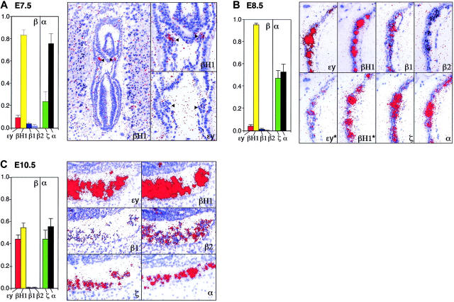
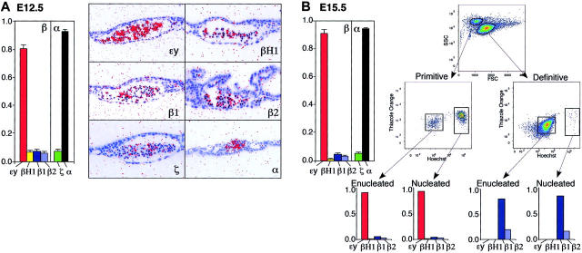
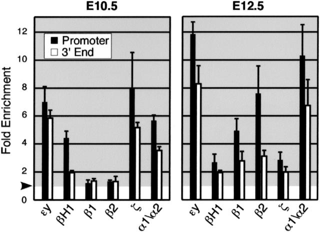
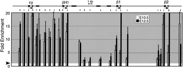
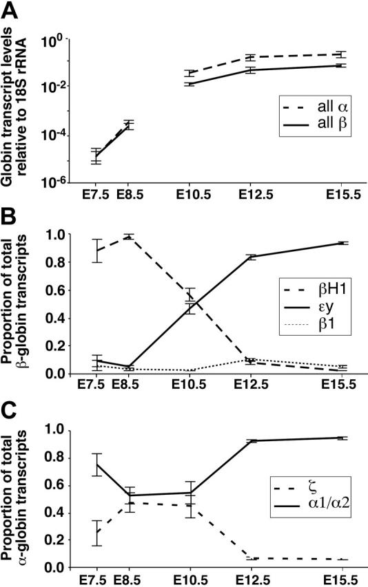
Mammals have 2 distinct erythroid lineages. The primitive erythroid lineage originates in the yolk sac and generates a cohort of large erythroblasts that terminally differentiate in the bloodstream. The definitive erythroid lineage generates smaller enucleated erythrocytes that become the predominant cell in fetal and postnatal circulation. These lineages also have distinct globin expression patterns. Our studies in primary murine primitive erythroid cells indicate that betaH1 is the predominant beta-globin transcript in the early yolk sac. Thus, unlike the human, murine beta-globin genes are not up-regulated in the order of their chromosomal arrangement. As primitive erythroblasts mature from proerythroblasts to reticulocytes, they undergo a betaH1- to epsilony-globin switch, up-regulate adult beta1- and beta2-globins, and down-regulate zeta-globin. These changes in transcript levels correlate with changes in RNA polymerase II density at their promoters and transcribed regions. Furthermore, the epsilony- and betaH1-globin genes in primitive erythroblasts reside within a single large hyperacetylated domain. These data suggest that this "maturational" betaH1- to epsilony-globin switch is dynamically regulated at the transcriptional level. Globin switching during ontogeny is due not only to the sequential appearance of primitive and definitive lineages but also to changes in globin expression as primitive erythroblasts mature in the bloodstream.


Figures

Figure 1.
Figure 1.
Changes in globin gene mRNA accumulation. (A) E7.5 yolk sac, (B) E8.5 yolk sac, and (C) E10.5 peripheral blood. Relative levels (mean ± SEM of at least 3 independent experiments) of the β-globin genes (εy, βH1, β1, and β2) and of the α-globin genes (ζ, α1/α2) quantified by qPCR are shown on the left. Total β- and total α-like globin transcript levels are each considered as 1. Expression of these same genes by in situ hybridization is shown in the right panels. Arrowheads indicate blood islands. For comparison, at each developmental time point, similar exposures of all of the globin genes are shown. In panel B, short exposures of the βH1- and εy-globins, marked by asterisks, highlight their differential transcript accumulation. ζ- and α1/α2-globin transcripts were not consistently detected at E7.5 (data not shown). Images were processed as described in “Materials and methods.”
Figure 2.
Figure 2.
Changes in globin gene mRNA accumulation in primitive erythroid cells. (A) E12.5 and (B) E15.5 of mouse gestation. Relative levels (mean ± SEM of at least 3 independent experiments) of the β-globin genes (εy, βH1, β1, and β2) and of the α-globin genes (ζ, α1/α2) quantified by qPCR from nucleated primitive erythroid cells are shown on the left. Total β- and total α-like globin transcript levels are each considered as 1. Globin transcript accumulation in circulating blood cells at E12.5 visualized by in situ hybridization is shown in the right panels of A. Isolation by FACS of primitive and definitive orthochromatic erythroblasts and reticulocytes from E15.5 peripheral blood is shown on the right panels in B. These populations can be distinguished by forward-side scatter and further isolated as Ter119-positive cells (not shown). Orthochromatic erythroblasts can be distinguished from reticulocytes after staining RNA with thiazole orange and DNA with Hoechst. Also shown are the patterns of β-globin gene expression in each purified population quantified by qPCR. Late-stage primitive erythroid cells at E15.5 express predominantly εy-globin transcripts, while late stage definitive erythroid cells express only β1- and β2-globin transcripts. Images were processed as described in “Materials and methods.”
Figure 3.
Figure 3.
RNA polymerase II association with α- and β-globin gene sequences in primitive erythroid cells. Enrichments are shown in bar graph format relative to multiple control sequences elsewhere in the β-globin locus, of crosslinked chromatin samples immunoprecipitated using antiserum against RNA polymerase II, as determined by quantitative real-time PCR. ▪ represent enrichments at the indicated β-globin gene promoters, while □ represent enrichments at sequences located within the third exon or immediately downstream of the stop codon for each gene, approximately 1 kb 3′ of the promoters. The border between shaded and unshaded regions represents relative enrichment of 1.0-fold (ie, no enrichment [arrowhead]). The data shown represent averages of at least 3 PCRs for each sequence examined, derived from 2 (E10.5) or 3 (E12.5) separate immunoprecipitations. Error bars represent SEM.
Figure 4.
Figure 4.
Histone modifications within the β-globin locus in primary primitive erythroid cells at E10.5 and E12.5 indicate that the εy- and βH1-globin genes are contained within a single, large, hyperacetylated domain. The β-globin locus is represented to scale at the top, with the active globin genes indicated by arrows and exons of these genes by the thickest portions of the line. Unlabeled, thicker lines represent the positions of β-globin pseudogenes. The positions of PCR-amplified regions analyzed in the ChIP assay are indicated immediately below. Shown in bar graph format are enrichments, relative to multiple control sequences derived from loci containing genes inactive in erythroid cells, of cross-linked chromatin samples immunoprecipitated using antiserum against histone H3 acetylated at lysines 9 and/or 16. Chromatin was isolated from peripheral blood at E10.5 (□) and E12.5 (▪). The border between shaded and unshaded regions in each graph represents relative enrichment of 1.0-fold (arrowhead). Values for enrichment greater than 20-fold have been truncated in this representation. Error bars represent SEM.
Figure 5.
Figure 5.
Summary of changes in relative globin mRNA levels, normalized to 18S rRNA, as primitive erythroid cells mature from proerythroblasts at E7.5 to E8.5 to reticulocytes at E15.5 in the mouse. The E7.5 and E8.5 values are based on dissected yolk sac tissues, E10.5 on whole peripheral blood, and E12.5 and E15.5 values on sorted cell populations. (A) Levels of total α-globin and β-globin gene transcripts quantified by qPCR suggest that total α-globin transcripts accumulate in excess of β-globin transcripts. (B) Relative levels of εy-, βH1-, and β1-globin transcripts reveal the βH1- to εy-globin switch between E8.5 and E12.5. (C) Relative levels of the ζ- and α1/α2-globin transcripts reveal the predominance of α1/α2-globin gene from the earliest stages of primitive erythroid maturation. Error bars represent SEM.

Similar articles

Cited by

References

    1. Wong PMC, Chung S-H, Reicheld SM, Chui DHK. Hemoglobin switching during murine embryonic development: evidence for two populations of embryonic erythropoietic progenitor cells. Blood. 1986;67: 716-721. - PubMed
    1. Palis J, Robertson S, Kennedy M, Wall C, Keller G. Development of erythroid and myeloid progenitors in the yolk sac and embryo proper of the mouse. Development. 1999;126: 5073-5084. - PubMed
    1. McGrath KE, Koniski AD, Malik J, Palis J. Circulation is established in a stepwise pattern in the mammalian embryo. Blood. 2003;101: 1669-1676. - PubMed
    1. de la Chapelle A, Fantoni A, Marks P. Differentiation of mammalian somatic cells: DNA and hemoglobin synthesis in fetal mouse yolk sac erythroid cells. Proc Natl Acad Sci U S A. 1969;63: 812-819. - PMC - PubMed
    1. Steiner R, Vogel H. On the kinetics of erythroid cell differentiation in fetal mice, I: microspectrophotometric determination of the hemoglobin content in erythroid cells during gestation. J Cell Physiol. 1973;81: 323-338. - PubMed

Publication types