. 2005 Nov 1;19(21):2593-605.
doi: 10.1101/gad.349305.

The PAS/LOV protein VIVID supports a rapidly dampened daytime oscillator that facilitates entrainment of the Neurospora circadian clock

Affiliations

The PAS/LOV protein VIVID supports a rapidly dampened daytime oscillator that facilitates entrainment of the Neurospora circadian clock

Mark Elvin et al. Genes Dev. .

Abstract

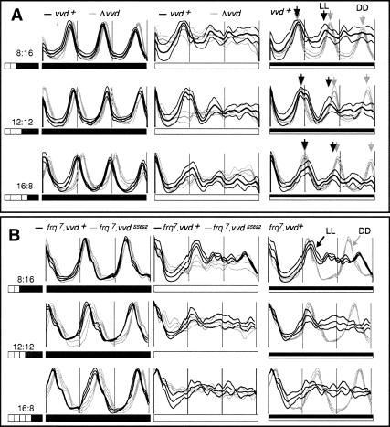
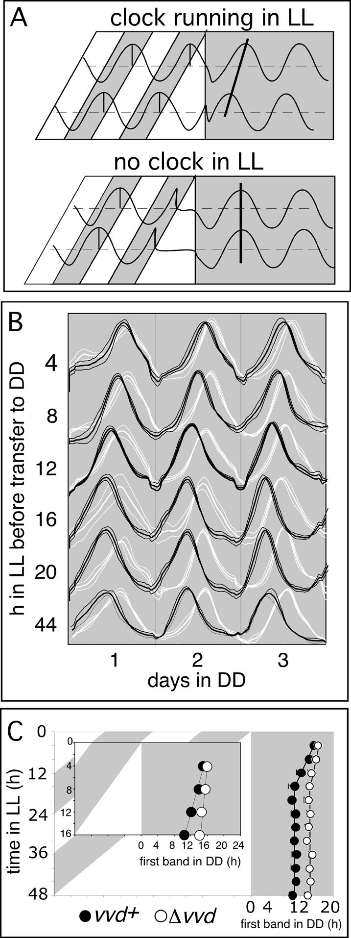
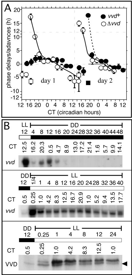
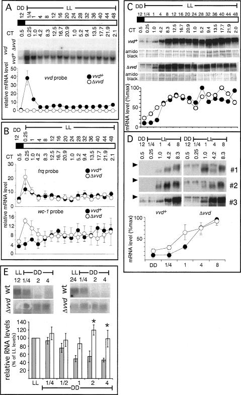
A light-entrainable circadian clock controls development and physiology in Neurospora crassa. Existing simple models for resetting based on light pulses (so-called nonparametric entrainment) predict that constant light should quickly send the clock to an arrhythmic state; however, such a clock would be of little use to an organism in changing photoperiods in the wild, and we confirm that true, albeit dampened, rhythmicity can be observed in extended light. This rhythmicity requires the PAS/LOV protein VIVID (VVD) that acts, in the light, to facilitate expression of an oscillator that is related to, but distinguishable from, the classic FREQUENCY/WHITE-COLLAR complex (FRQ/WCC)-based oscillator that runs in darkness. VVD prevents light resetting of the clock at dawn but, by influencing frq RNA turnover, promotes resetting at dusk, thereby allowing the clock to run through the dawn transition and take its phase cues from dusk. Consistent with this, loss of VVD yields a clock whose performance follows the simple predictions of earlier models, and overexpression of VVD restores rhythmicity in the light and sensitivity of phase to the duration of the photoperiod.


Figures

Figure 1.
Figure 1.
vvd permits a damped circadian clock to control conidiation in LL by preventing clock resetting at dawn. Averaged densitometric traces of 12-18 race tubes entrained for at least 2 d to photoperiods ranging from LD 8:16 to LD 16:8 before being released into DD (all left panels) or LL (all middle panels). The full set of investigated photoperiods are shown in Supplementary Figure S1. The right panels show DD and LL plots super-imposed with alignment at dusk (LL plots), and subjective dusk (DD plots), respectively. (A) Race tubes were inoculated with either wild-type (black traces) or vvdKO strains (gray traces). In the right panels, arrows indicate peaks of conidiation in wild type in DD (gray arrows) and LL (black arrows), respectively, to highlight coincident and diverging conidial rhythmicity. (B) As in A, but race tubes were inoculated with either frq7 (black traces) or frq7, vvdSS692 double mutants (gray traces). (Thick lines) Mean; (thin lines) ±1 SD; (white bars) light; (black bars) darkness.
Figure 2.
Figure 2.
vvd controls photoperiod-dependent changes in clock phase in DD. (A) Theoretical outcome of a release experiment revealing the existence (top panel) or absence (bottom panel) of clock function in LL. After entrainment to LD 12:12 cycles, Neurospora is released into LL of increasing lengths before its free-running rhythmicity is evaluated in DD. (Top panel) In the presence of a clock running in LL, the phase of the subsequent DD rhythm will depend on the phase of the LL clock at the dusk transition. Clock phase as measured in DD will systematically change depending on the previous day length. (Bottom panel) In the absence of an LL clock, the phase of the free-running rhythm in DD is independent of day length and is always set to the same phase at the dusk transition. (B) Averaged densitometric traces of wild-type (vvd+, black traces) and vvdKOvvd, white traces) strains cultured in a release experiment as depicted in A. (Thick lines) Mean; (thin lines) ±1 SD. (C) Phase of first conidial band in DD in vvdKOvvd, open circles) and wild-type (vvd+, filled circles) strains, as obtained in the race tube assays described in B. (Inset) Enlarged data set for race tubes released in up to 16 h LL.
Figure 3.
Figure 3.
vvd silences the resetting response to a standard light pulse during the first day in DD. (A) The light pulse PRC was obtained as described in Materials and Methods. The PRC is plotted in circadian hours, taking into account the 4-h phase difference between both strains. The center of the conidial band was defined as CT0; i.e., subjective dawn. The PRC is measured as the difference in the position of the conidial band with respect to unpulsed controls. (Abscissa) Circadian time (CT) in hours when light pulse was given; (ordinate) phase shift in hours of advance or delay; (filled circles) wild type (vvd+); (open circles) vvdKOvvd); (solid lines) trend lines for Δvvd; (dashed lines) trend lines vvd+ PRC; (error bars) ±SD; n = 6-18. (B) Northern and Western blot analysis of vvd RNA and VVD protein expression in DD and LL in wild type. Cultures were grown as described in experimental conditions, and RNA or protein was extracted from samples spanning 1-2 d in DD or LL. Sampling times are given both in real hours in DD or LL or CT. The top and middle panels show vvd expression profile in DD and LL, respectively. (Bottom) VVD protein levels in DD 12 and the following day in LL. Forty micrograms of total RNA and 100 μg of protein were loaded per lane. Northern blots were hybridized with a radiolabeled vvd probe and Western blots with an anti-serum directed against the VVD ORF. The black arrowhead indicates an unspecific hybridization signal, which is also present in vvdKO strains (data not shown). (White bars) Light; (black bars) darkness.
Figure 4.
Figure 4.
The classical FRQ/WCC oscillator stops early in LL, and vvd helps to set clock phase at dusk. (A,B) Northern blot and densitometric analysis of vvd, wc-1, and frq transcripts in wild-type (vvd+) or mutant (Δvvd) strains (n = 3). Cultures were subjected to various lengths of light (see Materials and Methods) before harvest and RNA or protein extraction. Controls were kept in DD. Forty micrograms of total RNA were loaded per lane. Transcript levels are given in arbitrary units. (Filled circles) Wild type; (open circles) vvdKO strains; (white bars) LL; (shaded bars) DD; (error bars) ±1 SE. (C, top panels) Representative Western blots (top) and their quantitation (bottom) showing FRQ accumulation in LL in wild type (vvd+) and vvdKOvvd). (D) Western blots (top three panels) and their quantitation (bottom) showing FRQ protein levels in the first 8 h in LL (three independent experiments) to emphasize differences in FRQ protein accumulation between mutant and wild type during the early light response. The arrowhead highlights the different FRQ form accumulating in vvdKOvvd). One-hundred micrograms of protein were loaded in each lane. (E) Northern analysis of frq expression levels in DD after exposure to different photoperiods (LL 12 and LL 24). Cultures were grown as described in Materials and Methods, and RNA was extracted from control samples before lights off (LL) and over a period of 4 h into subsequent DD. Northern blots were hybridized with a radiolabeled vvd probe. The densitometric analysis from three replicate experiments of a more detailed time-course is shown below. RNA levels are given relative to LL values, which were arbitrarily set to 100%. (Dark bars) Wild type (vvd+); (white bars) vvdKOvvd); (error bars) ±1 SE; (asterisks) differences that are statistically significant at the 0.05 confidence level.
Figure 5.
Figure 5.
Constitutive VVD expression rescues clock-phase and light-resetting defects in vvd knockout strains. (A, top) Generation of strains that express qa-2-inducible VVD. (Top) Plasmid pME11 contains a translational fusion of the qa-2 promoter (shaded dark gray) to the vvd ORF (hatched area), inserted into 5 kb of genomic his-3 region, which is available for homologous recombination. (Middle) Genomic organization of his-3 locus of parental strain 289-10 (Δvvd, bd, his). (Bottom) Genomic organization of qa-vvd knock-in strain created by homologous recombination at the his locus. Predicted SalI fragments are shown. (LG I) Linkage group I; (B) BamHI; (N) NotI; (P) PstI; (S) SalI; (arrows) direction of transcription. (B) Southern blot analysis of SalI-digested genomic DNA of primary transformants (pME11PT) and progeny from backcrosses to the Δvvd, his- parent (40-2 to 40-9). The arrow indicates the position of the expected genomic fragment containing the inserted vvd gene. (C) Phase of the first band of conidiation in DD of parental vvd knockout strain (Δvvd, open circles) and a representative qa-vvd knock-in strain (hatched circles) grown on increasing concentrations of the inducer quinic acid. Race tubes were grown for 2 d in LL before transfer to DD. Clock phase was analyzed for three independent experiments and plotted against quinic acid concentration. (Error bars) ±1 SD. (D) Phase response of wild type (vvd+, filled circles) vvdKOvvd, open circles), and a qa-vvd knock-in strain (hatched circles). The light pulse PRC was obtained as described in Figure 3A. (Abscissa) Circadian time (CT) in hours when light pulse was given; (ordinate) phase shift in hours of advance or delay. (E) Photoperiod-dependent phase is rescued in a qa-vvd knock-in strain. The experimental procedure is the same as described in Figure 2C. (Inset) Enlarged data set for race tubes released in up to 16 h LL.
Figure 6.
Figure 6.
VVD facilitates entrainment to continuous photoperiods. (A) Averaged densitometric traces of race tubes inoculated with wild type (87-3, black traces) or Δvvd (289-1, gray traces) grown in LD 12:12 entrainment conditions; n = 6-18. Plots are aligned at the dark-light boundaries. (Thick line) Mean; (thin lines) ±1 SD; (white bars) light; (black bars) darkness. Note the second peak of conidiation in the Δvvd strain. (B) Averaged densitometric traces of race tubes inoculated with wild-type (87-3, black traces) or Δvvd strains (289-1, gray traces) grown in skeleton or complete photoperiods. (Top panels) DD controls. (Second panels) 8:16 (left) and 16:8 (right) skeleton photoperiods. (Third panels) 8:16 (left) and 16:8 (right) complete photoperiods. Note the different phases of conidiation under skeleton and complete photoperiods in wild type in 8:16 photoperiods. Δvvd strains show similar phases of entrainment in both conditions. Also note that Δvvd strains always interpret the long dark interval between light pulses as night. (Bottom) LD 8:16 (left) and LD 16:8 (right) complete photoperiods, followed by corresponding skeleton photoperiods. Note the phase reversal of Δvvd strains that is not seen in wild type. For a more detailed discussion of this figure, see Results and Discussion.
Figure 7.
Figure 7.
A molecular model for the entrained Neurospora circadian clock. (A-E) For reasons of clarity, the Neurospora oscillator is depicted here as a simplified feedback loop built from the WCC and FRQ protein(s). For a limit cycle depiction of this figure, see Supplementary Figure S3. (A) In DD a robust circadian clock operates in wild-type (vvd+) and (Δvvd) knockout strains. (B) In wild type (vvd+) grown in LL, VVD represses light-input pathways and prevents light resetting of the oscillator, thus allowing for the temporary formation of a functional feedback loop in LL—however, with dampened amplitude—resulting in clear circadian conidiation cycles only for the first day or two. (C) In vvd knockout strains (Δvvd) growing in LL, the absence of VVD protein allows the WCC to reset the oscillator and override its negative feedback regulation by FRQ, effectively preventing the formation of a functional feedback loop. (D) In LD entrainment, a wild-type strain establishes an entrained oscillator consisting of segments of the previously described LL and DD oscillators. The phase of the oscillator at dusk, and thus the phase of circadian conidiation, is photoperiod dependent. (E) In LD entrainment, a vvd knockout strain (Δvvd) is reset at dawn and the oscillator resumes at dusk at a fixed and delayed phase compared with wild type. Clock phase in vvd knockout strains is largely independent of photoperiod. (Lines with arrowheads) Activating pathways; (blocked lines) inhibitory pathways; (dashed lines) partially functional pathways; (gray lines) light-signaling pathways.

Similar articles

Cited by

References

    1. Aronson B.D., Johnson, K.A., and Dunlap, J.C. 1994a. Circadian clock locus frequency: Protein encoded by a single open reading frame defines period length and temperature compensation. Proc. Natl. Acad. Sci. 91: 7683-7687. - PMC - PubMed
    1. Aronson B.D., Johnson, K.A., Loros, J.J., and Dunlap, J.C. 1994b. Negative feedback defining a circadian clock: Auto-regulation of the clock gene frequency. Science 263: 1578-1584. - PubMed
    1. Ashmore L.J. and Sehgal, A. 2003. A fly's eye view of circadian entrainment. J. Biol. Rhythms 18: 206-216. - PubMed
    1. Cermakian N. and Sassone-Corsi, P. 2000. Multilevel regulation of the circadian clock. Nat. Rev. Mol. Cell. Biol. 1: 59-67. - PubMed
    1. Chang B. and Nakashima, H. 1997. Effects of light-dark cycles on the circadian conidiation rhythm in Neurospora crassa. J. Plant. Res. 110: 449-453.

Publication types

MeSH terms

Substances

LinkOut - more resources