. 2006 Feb 15;394(Pt 1):115-24.
doi: 10.1042/BJ20051329.

Gene expression patterns and catalytic properties of UDP-D-glucose 4-epimerases from barley (Hordeum vulgare L.)

Affiliations

Gene expression patterns and catalytic properties of UDP-D-glucose 4-epimerases from barley (Hordeum vulgare L.)

Qisen Zhang et al. Biochem J. .

Abstract

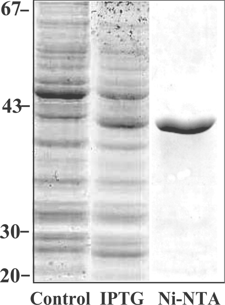
UGE (UDP-Glc 4-epimerase or UDP-Gal 4-epimerase; EC 5.1.3.2) catalyses the interconversion of UDP-Gal and UDP-Glc. Both nucleotide sugars act as activated sugar donors for the biosynthesis of cell wall polysaccharides such as cellulose, xyloglucans, (1,3;1,4)-beta-D-glucan and pectins, together with other biologically significant compounds including glycoproteins and glycolipids. Three members of the HvUGE (barley UGE) gene family, designated HvUGE1, HvUGE2 and HvUGE3, have been characterized. Q-PCR (quantitative real-time PCR) showed that HvUGE1 mRNA was most abundant in leaf tips and mature roots, but its expression levels were relatively low in basal leaves and root tips. The HvUGE2 gene was transcribed at significant levels in all organs examined, while HvUGE3 mRNA levels were very low in all the organs. Heterologous expression of a near full-length cDNA confirmed that HvUGE1 encodes a functional UGE. A non-covalently bound NAD+ was released from the enzyme after denaturing with aqueous ethanol and was identified by its spectrophotometric properties and by electrospray ionization MS. The K(m) values were 40 microM for UDP-Gal and 55 muM for UDP-Glc. HvUGE also catalyses the interconversion of UDP-GalNAc and UDP-GlcNAc, although it is not known if this has any biological significance. A three-dimensional model of the HvUGE revealed that its overall structural fold is highly conserved compared with the human UGE and provides a structural rationale for its ability to bind UDP-GlcNAc.


Figures

Figure 1
Figure 1. Alignment of amino acid sequences of UGEs
Amino acid sequences of UGEs from barley (HvUGE1, HvUGE2 and HvUGE3), A. thaliana (AtUGE1, Swiss-Prot Data Bank entry Q42605), rice (TIGR locus: Os05g51670) and human (PDB entry 1hzj) were aligned using the GeneDoc program (www.psc.edu/biomed/genedoc). The conserved motifs GXXGXXG (NAD+-binding) and catalytic amino acid residues Ser and Tyr-Xaa-Xaa-Xaa-Lys of the active site are underlined.
Figure 2
Figure 2. SDS/PAGE of barley HvUGE1 expressed in E. coli
The barley HvUGE1 cDNA was expressed in E. coli and proteins were separated by SDS/PAGE. Control, cell extracts without addition of IPTG; IPTG, protein expression following induction with 0.5 mM IPTG; Ni-NTA, the purified fraction following Ni-NTA affinity chromatography. Protein molecular mass standards (in kDa) are indicated on the left.
Figure 3
Figure 3. Normalized expression levels of HvUGE genes in different barley organs
Levels of mRNA are presented as the number of copies per μg of total RNA after normalization. Organs were as described by Burton et al. [16] and included leaf tips, leaf bases, root tip and the maturation zone (m/zone) of roots, flowers just before anthesis (FL early), flowers at anthesis (FL anthesis), developing grain 3 and 13 days post-anthesis (Grain early and Grain mid), 3-day-old coleoptiles (Col 3 day) and the stem of mature plants just below the emerging ear (Stem). The results shown are means for four separate evaluations and S.D.s are indicated.
Figure 4
Figure 4. Normalized expression levels of HvUGE genes in young barley leaves, roots and coleoptiles
Barley leaves were sectioned into five segments and roots were cut into four segments as described by Burton et al. [16]. Units and statistical treatments were as described in the legend to Figure 3.
Figure 5
Figure 5. Molecular model of the HvUGE1
(A) Stereo view of the ribbon representation of the HvUGE1 model shows secondary structural elements of the enzyme folded into a mixed α/β protein fold. The R.M.S.D. value between the template structure (PDB entry 1hzj) and the modelled HvUGE1 is 0.951 Å over 343 amino acid residues. The N- and C-termini of the enzyme are shown in the lower and upper parts of the panel respectively. Spheres in atomic colours represent van der Waals radii of bound UDP-GlcNAc and NAD+ ligands. (B) Stereo representation of the molecular surface area depicts the active site region with bound NAD+ and UDP-GlcNAc. The molecular surface was generated with a probe radius of 1.4 Å. The Figure was prepared with PyMol.

Similar articles

Cited by

References

    1. Reiter W. D., Vanzin G. F. Molecular genetics of nucleotide sugar interconversion pathways in plants. Plant Mol. Biol. 2001;47:95–113. - PubMed
    1. Seifert G. J. Nucleotide sugar interconversions and cell wall biosynthesis: how to bring the inside to the outside. Curr. Opin. Plant Biol. 2004;7:277–284. - PubMed
    1. Zhang Q., Shirley N., Lahnstein J., Fincher G. B. Characterization and expression patterns of UDP-D-glucuronate decarboxylase genes in barley. Plant Physiol. 2005;138:131–141. - PMC - PubMed
    1. Bauer A. J., Rayment I., Frey P. A., Holden H. M. The molecular structure of UDP-galactose 4-epimerase from Escherichia coli determined at 2.5 Å resolution. Proteins. 1992;12:372–381. - PubMed
    1. Barat B., Bhattacharyya D. UDP-galactose 4-epimerase from Escherichia coli: formation of catalytic site during reversible folding. Arch. Biochem. Biophys. 2001;391:188–196. - PubMed

Publication types

MeSH terms

Substances

Associated data

LinkOut - more resources