Comparative Study
. 2005 Nov 2;25(44):10247-51.
doi: 10.1523/JNEUROSCI.1089-05.2005.

Neuronal L-type calcium channels open quickly and are inhibited slowly

Affiliations
Comparative Study

Neuronal L-type calcium channels open quickly and are inhibited slowly

Thomas D Helton et al. J Neurosci. .

Abstract

Neuronal L-type calcium channels are essential for regulating activity-dependent gene expression, but they are thought to open too slowly to contribute to action potential-dependent calcium entry. A complication of studying native L-type channels is that they represent a minor fraction of the whole-cell calcium current in most neurons. Dihydropyridine antagonists are therefore widely used to establish the contribution of L-type channels to various neuronal processes and to study their underlying biophysical properties. The effectiveness of these antagonists on L-type channels, however, varies with stimulus and channel subtype. Here, we study recombinant neuronal L-type calcium channels, CaV1.2 and CaV1.3. We show that these channels open with fast kinetics and carry substantial calcium entry in response to individual action potential waveforms, contrary to most studies of native L-type currents. Neuronal CaV1.3 L-type channels were as efficient as CaV2.2 N-type channels at supporting calcium entry during action potential-like stimuli. We conclude that the apparent slow activation of native L-type currents and their lack of contribution to single action potentials reflect the state-dependent nature of the dihydropyridine antagonists used to study them, not the underlying properties of L-type channels.


Figures

Figure 1.
Figure 1.
Neuronal L-type channels activate over a wide voltage range and with fast kinetics. a, Normalized, averaged, current-voltage relationships for calcium currents recorded from tsA201 cells expressing CaV1.3 (▪), CaV1.2 (•), CaV2.2 (▴), and CaV3.1 (♦) subunit cDNAs. CaVα was coexpressed with CaVβ3 and CaVα2δ1 cDNAs. Average peak current amplitudes were the following: CaV1.3, -1.7 ± 0.4 nA (n = 12); CaV1.2, -0.5 ± 0.04 nA (n = 8); CaV2.2, -1.9 ± 0.3 nA (n = 6); CaV3.1, -1.3 ± 0.2 nA (n = 8). Activation midpoints (in millivolts) estimated from Boltzmann-GHK fits of data were the following: CaV1.3, -39.4 ± 0.6 mV (n = 8); CaV1.2, -17.6 ± 0.7 mV (n = 11); CaV2.2, -12.7 ± 0.8 mV (n = 8); and CaV3.1, -46.9 ± 1.2 mV (n = 8). b, Normalized representative current traces for CaV1.2, CaV1.3, CaV2.2 (gray trace), and CaV3.1 (gray trace) activated by step depolarization to activation midpoints: CaV1.3, -40 mV; CaV1.2, -15 mV; CaV2.2, -15 mV; and CaV3.1, -45 mV. c, Averaged macroscopic activation time constants (ln τact) at different test potentials estimated from exponential fits to currents recorded from cells expressing CaV1.2, CaV1.3, CaV2.2, and CaV3.1. Values are mean ± SE. The lines show regression fits to the data. Slopes and y intercepts are the following: CaV1.3, -0.02 ± 0.001 mV-1, 0.23 ± 0.02 (n = 9); CaV1.2, -0.02 ± 0.003 mV-1, 0.89 ± 0.09 (n = 11); CaV2.2, -0.02 ± 0.001 mV-1, -0.11 ± 0.01 (n = 7); and CaV3.1, -0.049 ± 5 × 10⁻⁴ mV-1, -0.63 ± 0.07 (n = 6). Student's t test on time constants at all test potentials: CaV1.3 to CaV2.2, p > 0.27; CaV1.3 to CaV1.2, p < 0.001.
Figure 2.
Figure 2.
CaV1.3 activates rapidly in response to action potential-like (AP) waveforms. Overlaid normalized (a) and non-normalized (b) representative current traces for CaV1.2, CaV1.3, and CaV2.2 channels in response to AP waveform are shown. The AP was recorded from a sympathetic neuron and was triggered by a brief current injection seen as a hump on the foot of the waveform. c, Average time delay between the peak of the action potential waveform (APPK) and peak inward current (IPK). Time delays were the following: CaV1.3, 0.65 ± 0.06 ms (n = 11); CaV1.2, 0.81 ± 0.05 ms (n = 8); and CaV2.2, 0.63 ± 0.05 ms (n = 8). CaV1.3 and CaV2.2 values were not significantly different; CaV1.2 and CaV1.3 values were significantly different (*p < 0.05). d, Averaged ratios of total charge moved during a single AP to peak inward current evoked from a 50 ms step depolarization. Average values were the following: CaV1.3, 0.84 ± 0.06 (n = 19); CaV1.2, 0.61 ± 0.05 (n = 8); and CaV2.2, 0.94 ± 0.11 (n = 6). Average AP peak currents for CaV1.2, CaV1.3, and CaV2.2 were 663 ± 137 pA (n = 8), 1282 ± 110 pA (n = 19), and 2898 ± 1273 pA (n = 10), respectively. IV, Current-voltage. Error bars represent SE.
Figure 3.
Figure 3.
Nifedipine inhibits CaV1.2 and CaV1.3 channels activated by step depolarization. The time courses of inhibition of CaV1.2 (a; •) and CaV1.3 (b; ▪) currents by 5 μm nifedipine (n = 7 and n = 8, respectively) are shown. Currents were evoked by 50 ms square pulse depolarizations to 0 mV (CaV1.2) and -20 mV (CaV1.3) from a holding potential of -80 mV. Depolarizations were applied once every 2 s. The bars show duration of nifedipine application. Insets, Representative CaV1.2 and CaV1.3 currents before (Con) and after exposure to nifedipine (Nif; 16 s time point). Calibration: 0.2 nA, 10 ms for CaV1.2 (a) and 0.5 nA, 10 ms for CaV1.3 (b). Error bars represent SE.
Figure 4.
Figure 4.
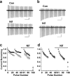
Nifedipine is weakly effective on CaV1.2 and CaV1.3 channels activated by action potential-like stimuli. Representative CaV1.2 (a) and CaV1.3 (b) currents evoked by a train of action potential waveforms, applied at 100 Hz from holding potentials of -80 mV, before (Con) and after a 10 s exposure to 5 μm nifedipine (Nif), are shown. Calibration: a, 0.2 nA, 50 ms; b, 0.5 nA, 50 ms. The action potential waveform used as command voltage was recorded from a sympathetic neuron and triggered by a brief current injection. Average peak current amplitudes for CaV1.2 (c; •, ○) and CaV1.3 (d; ▪, □) measured from currents induced by trains of 30 action potentials before and 10 s after exposure to 5 μm nifedipine (Nif) are shown. Currents were evoked from holding potentials of -80 mV (•, ▪) and -60 mV (○, □). Current amplitudes at the end of a series of four stimulus trains in the presence of nifedipine are shown for each series (180th pulse). Currents recovered completely after removal of nifedipine within three stimulus trains. Recovery was slowed approximately three fold when the membrane potential was depolarized to -60 mV. Error bars represent SE.

References

    1. Avery RB, Johnston D (1996) Multiple channel types contribute to the low-voltage-activated calcium current in hippocampal CA3 pyramidal neurons. J Neurosci 16: 5567-5582. - PMC - PubMed
    1. Bean BP (1984) Nitrendipine block of cardiac calcium channels: high-affinity binding to the inactivated state. Proc Natl Acad Sci USA 81: 6388-6392. - PMC - PubMed
    1. Berjukow S, Hering S (2001) Voltage-dependent acceleration of Ca(v)1.2 channel current decay by (+)- and (-)-isradipine. Br J Pharmacol 133: 959-966. - PMC - PubMed
    1. Birnbaumer L, Qin N, Olcese R, Tareilus E, Platano D, Costantin J, Stefani E (1998) Structures and functions of calcium channel beta subunits. J Bioenerg Biomembr 30: 357-375. - PubMed
    1. Bonci A, Grillner P, Mercuri NB, Bernardi G (1998) L-type calcium channels mediate a slow excitatory synaptic transmission in rat midbrain dopaminergic neurons. J Neurosci 18: 6693-6703. - PMC - PubMed

Publication types