. 2006 Jan;17(1):336-44.
doi: 10.1091/mbc.e05-09-0874. Epub 2005 Nov 2.

CAR-1, a protein that localizes with the mRNA decapping component DCAP-1, is required for cytokinesis and ER organization in Caenorhabditis elegans embryos

Affiliations

CAR-1, a protein that localizes with the mRNA decapping component DCAP-1, is required for cytokinesis and ER organization in Caenorhabditis elegans embryos

Jayne M Squirrell et al. Mol Biol Cell. 2006 Jan.

Abstract

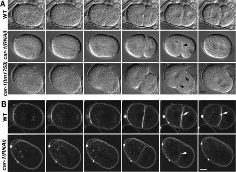
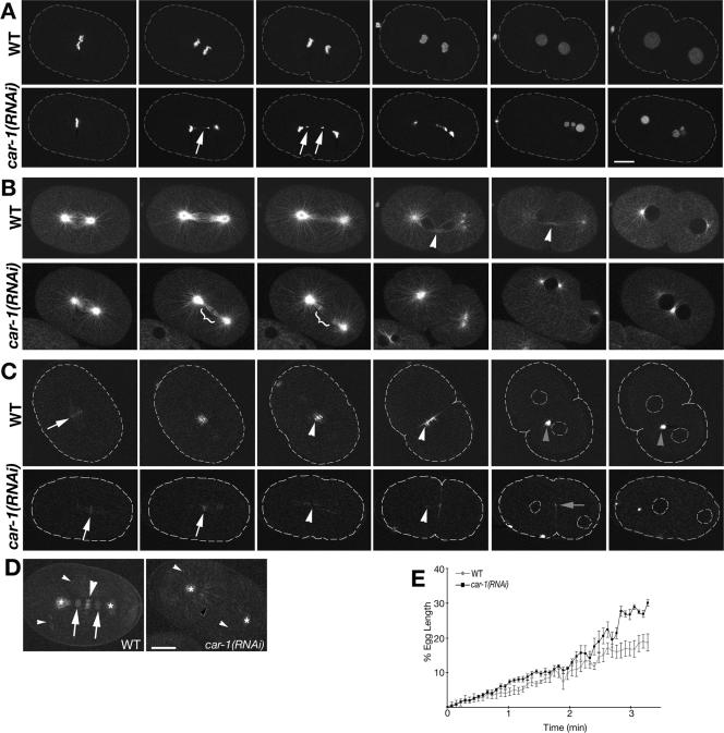
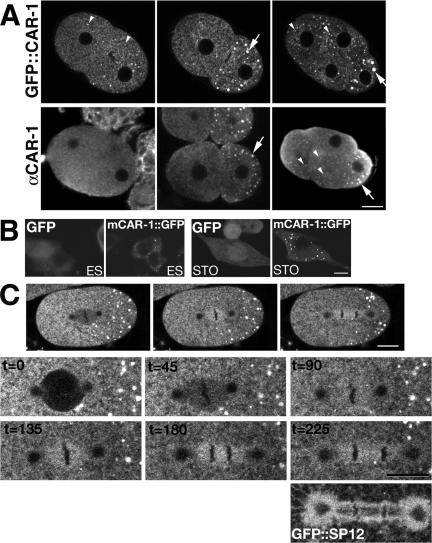
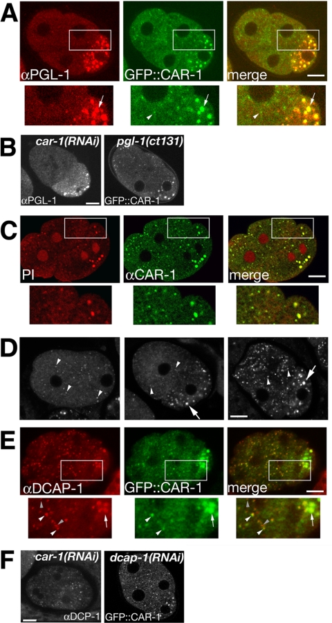
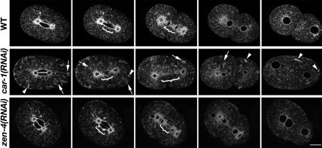
The division of one cell into two requires the coordination of multiple components. We describe a gene, car-1, whose product may provide a link between disparate cellular processes. Inhibition of car-1 expression in Caenorhabditis elegans embryos causes late cytokinesis failures: cleavage furrows ingress but subsequently regress and the spindle midzone fails to form, even though midzone components are present. The localized accumulation of membrane that normally develops at the apex of the cleavage furrow during the final phase of cytokinesis does not occur and organization of the endoplasmic reticulum is aberrant, indicative of a disruption in membrane trafficking. The car-1 gene has homologues in a number of species, including proteins that associate with RNA binding proteins. CAR-1 localizes to P-granules (germ-line specific ribonucleoprotein particles) and discrete, developmentally regulated cytoplasmic foci. These foci also contain DCAP-1, a protein involved in decapping mRNAs. Thus, CAR-1, a protein likely to be associated with RNA metabolism, plays an essential role in the late stage of cytokinesis, suggesting a novel link between RNA, membrane trafficking and cytokinesis in the C. elegans embryo.


Figures

Figure 1.
Figure 1.
CAR-1 depleted embryos fail late cytokinesis. Each time sequence illustrates the following developmental stages, from left to right: spindle formation; spindle elongation; further spindle elongation/cleavage furrow initiation; cleavage furrow completion; early 2-cell/cytokinesis failure; late 2 cell. (A) Top row is a wild-type embryo. Nomarski images showing that the depletion of CAR-1, either by RNAi (middle row) or a deletion mutation (car-1(tm1735)) (bottom row) caused the cleavage furrow to regress (black arrowhead) after it had fully ingressed. Asterisk denotes extra micronucleus in RNAi treated embryo. (B) In the wild-type embryo (top row) labeled with the membrane dye FM 2-10, there was a striking accumulation of membrane, after the complete ingression of the cleavage furrow, at the scission site (white arrow), which persisted through much of the cell cycle; no such accumulation was seen in the car-1(RNAi) embryo (bottom row). Instead, the membrane regressed (white arrowhead). For this and all other embryo figures, anterior is toward the left. Scale bar, 10 μm.
Figure 2.
Figure 2.
CAR-1 depleted embryos exhibit spindle midzone defects. Each multiphoton time sequence (A-C) illustrates the following developmental stages, from left to right: spindle formation; spindle elongation; further spindle elongation/cleavage furrow initiation; cleavage furrow completion; early 2-cell/cytokinesis failure; late 2 cell. For each series, top row is a wild-type embryo and the bottom row is a car-1(RNAi) embryo. (A) GFP::Histone expressing embryos treated with car-1(RNAi) exhibited lagging chromosome fragments during spindle elongation (white arrow), resulting in micronuclei. (B) In the control GFP::tubulin expressing embryo a prominent and persistent midbody was observed as the cleavage furrow completed (white arrowhead), which was absent in the car-1(RNAi) embryo. Also the spindle microtubule bundle became radially compressed compared with the control embryo during early elongation, giving the appearance of a thinner spindle, which seemed to ultimately break (bracket). (C) GFP::ZEN-4 localized to metaphase chromosomes in the car-1(RNAi) embryo, as it did in the control (white arrow). ZEN-4 moved to midzone microtubules in the RNAi treated embryo, as in the control (white arrowhead), but did not properly organize into a midbody structure (gray arrowhead in wild-type embryo). However, it did localize to the cleavage furrow, even as the cleavage furrow regressed (gray arrow). Bright spot in last image is an internalized remnant from the meiotic division. Dashed lines for GFP::ZEN4 and GFP::Histone sequences are embryo outlines taken from the corresponding brightfield images (unpublished data). (D) These images are projections of 10 time points (4 s apart) showing the localization SPD-1 during spindle elongation. In the control embryo, SPD-1 localized to the reforming nuclei (white arrows), the spindle midzone (large white arrowhead), centrosomes (asterisk), and cytoplasmic microtubules (small white arrowhead). car-1(RNAi) embryo showed the same localizations (nuclei not shown in this plane) except the microtubule labeling in the midzone region was more dispersed (small black arrowhead), resembling the cytoplasmic microtubule labeling. Scale bars, 10 μm. (E) The average change in spindle length is illustrated, in wild-type and in car-1(RNAi) embryos, measured as a percentage of egg length, showing that a greater change in the spindle length in the car-1(RNAi) embryos than in the untreated embryos, particularly later in elongation. The points are averages of three untreated or four car-1(RNAi) embryos expressing GFP::tubulin, with SEM shown for each point. Time 0 is the initiation of chromosome separation and measurements were taken at 4.37-s intervals.
Figure 3.
Figure 3.
CAR-1 localizes to cytoplasmic puncta and can associate with the anaphase spindle. (A) Columns indicate the following developmental stages: pronuclear migration, 2-cell interphase, and 4-cell interphase. The top row is a multiphoton time sequence of a living embryo showing changes in distribution of GFP::CAR-1, with few small cytoplasmic puncta (small arrowhead) present during pronuclear migration and larger puncta developing in the posterior during and after cytokinesis (arrow). There was an increase in the number of small cytoplasmic puncta during and after the 2-4 cell division; midpronuclear migration: 0.007 ± 0.004 puncta/μ2, N = 4 embryos; 2-cell interphase: 0.004 ± 0.003 puncta/μ2,N = 8 embryos; AB furrow initiation: 0.013 ± 0.004 puncta/μ2, N = 8 embryos; interphase Aba = 0.038 ± 0.007. An antibody to CAR-1 showed a similar pattern in fixed embryos (bottom row). (B) Confocal images of mouse embryonic stem cells (ES) or fibroblasts (STO) transfected with either GFP only or with the full-length mCAR-1 tagged with GFP. The mCAR-1::GFP was expressed in numerous discrete foci in both embryonic stem cells and in fibro-blasts. (C) Multiphoton time series of living embryos showing CAR-1 spindle localization. GFP::CAR-1 was initially absent from the spindle and then accumulated on the spindle region, and as the spindle elongated, the CAR-1 label extended along the spindle to include the pericentriolar region. Lower series shows an enlargement of the spindle region at 45-s intervals, with the final image compared with the organization of the ER (lowest image shows GFP::SP12). These images were taken with higher detector gain than those in A to accentuate the nonpunctate cytoplasmic distribution of GFP::CAR-1. Scale bars, 10 μm.
Figure 4.
Figure 4.
CAR-1 localizes with P-granules and particles containing RNA and DCAP-1. (A) Confocal images showing that the large puncta (white arrows) of GFP::CAR-1 (green) colocalized with the P-granule component PGL-1 (red), whereas the small puncta (white arrowhead) did not. (B) PGL-1 was present in CAR-1-depleted embryos and, conversely, GFP::CAR-1 was not disrupted in the pgl-1 mutant. Designations in bold at the top of each image indicate genotype while designation at bottom of each image indicate label. (C) Multiphoton microscopy images showing that both small and large CAR-1 punta (green) label with propidium iodide (PI; red). (D) Confocal micrographs showing the localization of DCAP-1 in large and small cytoplasmic puncta (top row: pronuclear migration, 2-cell interphase, 4-cell interphase). (E) Both the large (white arrow) and small (white arrowhead) CAR-1 puncta (green) colocalized with DCAP-1-positive puncta (red); however, some DCAP-1 puncta appear to lack clear CAR-1 signal (gray arrowhead). (F) DCAP-1 was present when CAR-1 was depleted by RNAi, and GFP::CAR-1 expression was present in dcap-1(RNAi) embryos. Scale bars, 10 μm.
Figure 5.
Figure 5.
CAR-1 depletion disrupts the organization of the ER. (A) Multiphoton time series of living embryos showing ER dynamics. Sequences illustrate the organization of the ER (visualized by GFP::SP12; Poteryaev et al., 2005) during premetaphase, metaphase, anaphase, telophase, and interphase (2-cell stage). In the control embryo (top row), the ER outlined the spindle and exhibited a distinct reticulate structure. This structure became dispersed as the cleavage furrow ingressed and the cell entered interphase. CAR-1 depletion (second row) perturbed ER reticulation during mitosis, causing patchy accumulations (arrows) or thick strands (arrowheads) of ER that persisted into interphase. The car-1(RNAi) embryo also showed a reduction in spindle associated ER in the midzone region (bracket) during mitosis progression. Although the zen-4(RNAi) embryo did have somewhat reduced midzone ER during anaphase, unlike the car-1(RNAi) embryo, substantial ER was still present near the reforming nuclei (bracket, middle panel) and the ER formed normal looking reticulate structures during metaphase. Scale bar, 10 μm.

Similar articles

Cited by

References

    1. Albrecht, M., and Lengauer, T. (2004). Novel Sm-like proteins with long C-terminal tails and associated methyltransferases. FEBS Lett. 569, 18-26. - PubMed
    1. Badrinath, A. S., and White, J. G. (2003). Contrasting patterns of mitochondrial redistribution in the early lineages of Caenorhabditis elegans and Acrobeloides sp. PS1146. Dev. Biol. 258, 70-75. - PubMed
    1. Bashirullah, A., Cooperstock, R. L., and Lipshitz, H. D. (2001). Spatial and temporal control of RNA stability. Proc. Natl. Acad. Sci. USA 98, 7025-7028. - PMC - PubMed
    1. Blower, M. D., Nachury, M., Heald, R., and Weis, K. (2005). A Rae1-containing ribonucleoprotein complex is required for mitotic spindle assembly. Cell 121, 223-234. - PubMed
    1. Boag, P., Nakamura, A., and Blackwell, T. K. (2005). A conserved RNA-protein complex component involved in physiological germline apoptosis regulation in C. elegans. Development 132, 4975-4986. - PubMed

Publication types

MeSH terms

LinkOut - more resources