. 2005 Nov 3;438(7064):94-8.
doi: 10.1038/nature04110.

The transcription factor Engrailed-2 guides retinal axons

Affiliations

The transcription factor Engrailed-2 guides retinal axons

Isabelle Brunet et al. Nature. .

Abstract

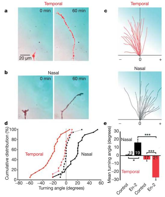
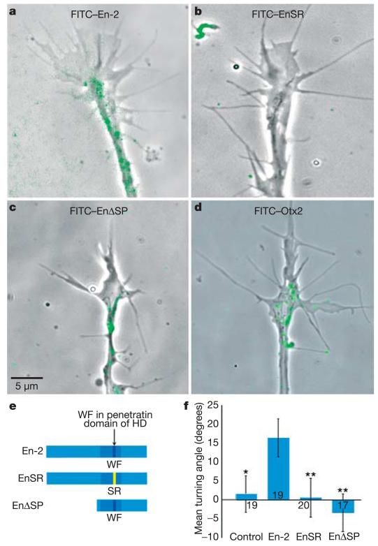
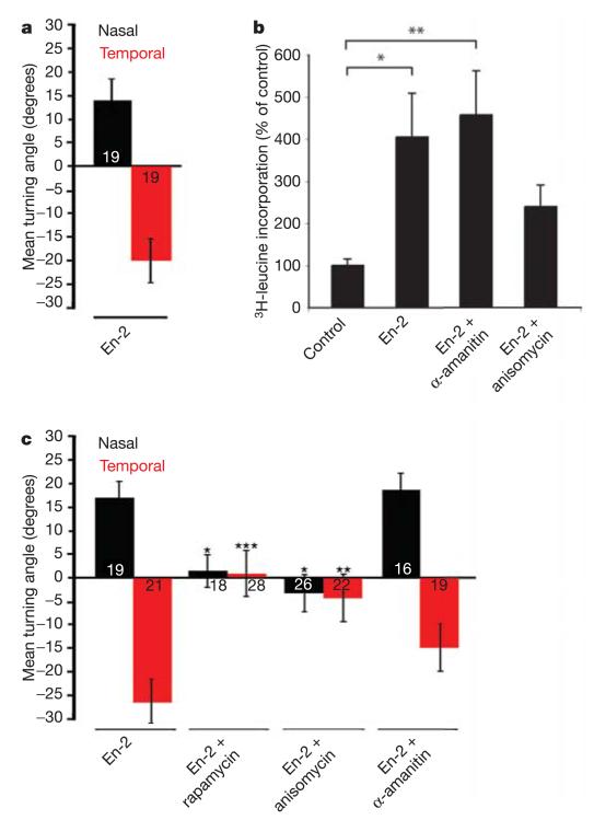
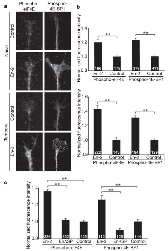
Engrailed-2 (En-2), a homeodomain transcription factor, is expressed in a caudal-to-rostral gradient in the developing midbrain, where it has an instructive role in patterning the optic tectum--the target of topographic retinal input. In addition to its well-known role in regulating gene expression through its DNA-binding domain, En-2 may also have a role in cell-cell communication, as suggested by the presence of other domains involved in nuclear export, secretion and internalization. Consistent with this possibility, here we report that an external gradient of En-2 protein strongly repels growth cones of Xenopus axons originating from the temporal retina and, conversely, attracts nasal axons. Fluorescently tagged En-2 accumulates inside growth cones within minutes of exposure, and a mutant form of the protein that cannot enter cells fails to elicit axon turning. Once internalized, En-2 stimulates the rapid phosphorylation of proteins involved in translation initiation and triggers the local synthesis of new proteins. Furthermore, the turning responses of both nasal and temporal growth cones in the presence of En-2 are blocked by inhibitors of protein synthesis. The differential guidance of nasal and temporal axons reported here suggests that En-2 may participate directly in topographic map formation in the vertebrate visual system.


Figures

Figure 1
Figure 1. En-2 gradient repels temporal and attracts nasal retinal axons
a, b, Examples of temporal (red) and nasal (black) growth cones tested in turning assays with 10 μg ml−1 En-2 in the pipette (300 pM at the growth cone). The pipette is positioned in the top right, and the En-2 gradient is represented in blue (ac). c, Trajectory plots of temporal (red) and nasal (black) neurites in En-2 gradients. Each line represents a single growth cone trajectory; the origin represents the centre of the growth cone at 0 min, and positive (+) and negative (−) turning angles are indicated. d, Cumulative distributions of turning angles of temporal (red) and nasal (black) growth cones in En-2 (bold) and control (light) gradients. e, Mean turning angles of growth cones in the experimental conditions shown in d, numbers on or beside the bars denote the number of growth cones tested. Significance was calculated using a Kolmogorov–Smirnov test, and indicated by asterisks (*P < 0.05; **P < 0.01; ***P < 0.001). Error bars indicate s.e.m.
Figure 2
Figure 2. En-2 is internalized in the growth cone and guidance depends on internalization
ac, Live retinal growth cones following 5 min exposure to FITC-tagged proteins. Over 50% of growth cones showed internalized fluorescent puncta (green) following exposure to FITC-En-2 (a) or FITC–EnΔSP (c), but not with FITC–EnSR (b). d, FITC–Otx2 is internalized. e, En-2 constructs used in the turning assays: En-2 is the full-length wild-type protein; EnSR has a mutation in the penetratin domain (amino acids WF replaced by SR); and EnΔSP lacks the N-terminal part of the protein (where a putative eIF4E-binding site resides). HD, homeodomain. f, Mean turning angles of nasal growth cones in gradients of En-2, EnSR and EnΔSP. Nasal axons turn towards En-2 but not EnSR or EnΔSP. For Otx2 activity, see Supplementary Fig. 1d. For P values, see Fig. 1 legend, and comparisons are to En-2. Error bars indicate s.e.m.
Figure 3
Figure 3. Retinal ganglion cell growth cone guidance by En-2 is dependent on protein synthesis
a, Temporal (red) and nasal (black) growth cones isolated from their somas (see Supplementary Fig. 1c) show opposite turning responses to an En-2 gradient, eliminating a role for transcription. b, 3H-leucine incorporation in growth cones isolated from their cell bodies. En-2 stimulates significant incorporation of 3H-leucine, which is abolished by anisomycin but not α-amanitin. c, Mean turning angles of temporal and nasal growth cones in a gradient of En-2 in the presence of inhibitors. En-2-induced turning is blocked by anisomycin and rapamycin, but is unaffected by α-amanitin. The number of growth cones tested in each case is indicated on the bars. b, Significance was calculated using a Kruskal–Wallis test, and indicated by asterisks (*P < 0.05; **P < 0.01). c, Significance was calculated using a Kolmogorov–Smirnov test, and indicated by asterisks (*P < 0.05; **P < 0.01; ***P < 0.001), and comparisons are to En-2. Error bars indicate s.e.m.
Figure 4
Figure 4. En-2 stimulates the phosphorylation of translational regulatory proteins
a, Fluorescent micrographs of unstimulated control and En-2-stimulated growth cones using phospho-eIF4E and phospho-eIF4E-BP1 antibodies. Exposure to En-2 for 5 min increases the level of phosphorylation of eIF4E and eIF4E-BP1 in nasal and temporal growth cones. b, Histograms showing the digital quantification of the fluorescence signal in growth cones. c, Exposure of growth cones to EnΔSP does not lead to phosphorylation of either eIF4E or eIF4E-BP1. The number of growth cones tested in each case is indicated on the bars. Significance was calculated using a two-tailed Mann–Whitney test, and indicated by asterisks (**P < 0.0001). Error bars indicate s.e.m.

References

    1. Retaux S, Harris WA. Engrailed and retinotectal topography. Trends Neurosci. 1996;19:542–546. - PubMed
    1. Itasaki N, Nakamura H. A role for gradient en expression in positional specification on the optic tectum. Neuron. 1996;16:55–62. - PubMed
    1. Prochiantz A, Joliot A. Can transcription factors function as cell-cell signalling molecules? Nature Rev. Mol. Cell Biol. 2003;4:814–819. - PubMed
    1. Sperry RW. Chemoaffinity in the orderly growth of nerve fiber patterns and connections. Proc. Natl Acad. Sci. USA. 1963;50:703–710. - PMC - PubMed
    1. Cheng HJ, Nakamoto M, Bergemann AD, Flanagan JG. Complementary gradients in expression and binding of ELF-1 and Mek4 in development of the topographic retinotectal projection map. Cell. 1995;82:371–381. - PubMed

Publication types

MeSH terms