Comparative Study
. 2005 Nov;17(11):1679-90.
doi: 10.1162/089892905774589208.

Delay-period activity in the prefrontal cortex: one function is sensory gating

Affiliations
Comparative Study

Delay-period activity in the prefrontal cortex: one function is sensory gating

Bradley R Postle. J Cogn Neurosci. 2005 Nov.

Abstract

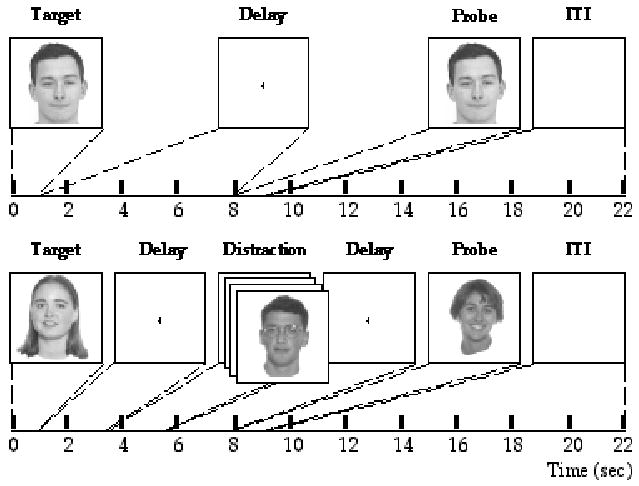
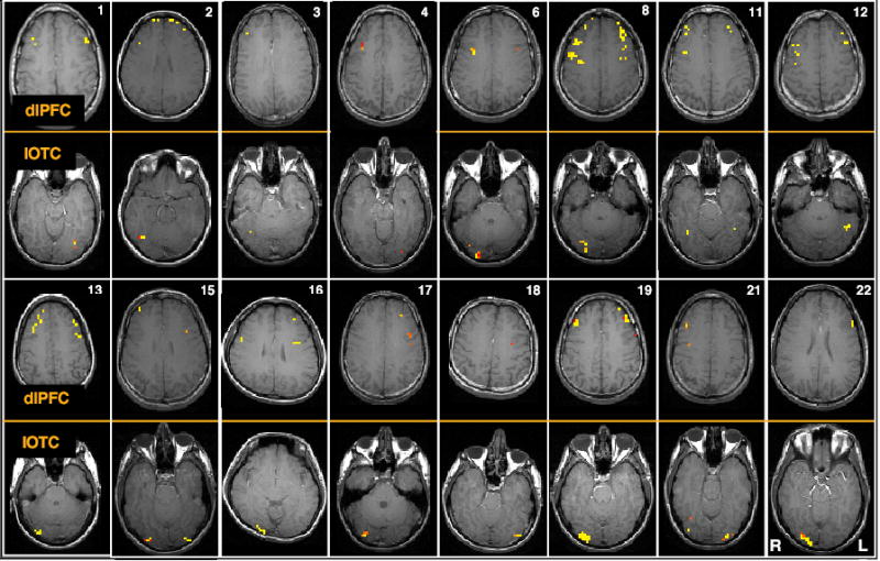
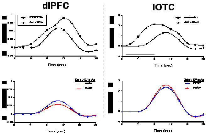
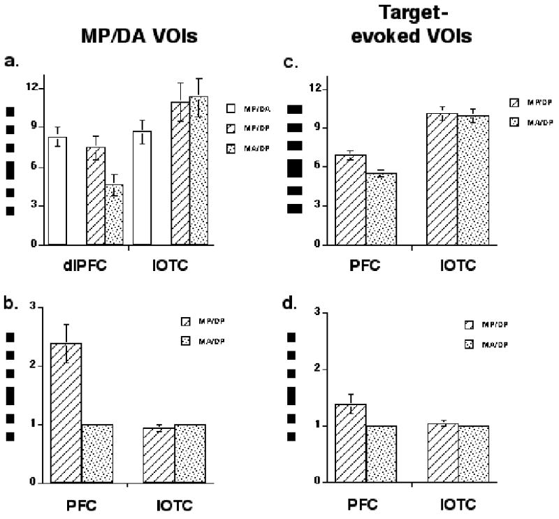
The prefrontal cortex (PFC) contributes to working memory functions via executive control processes that do not entail the storage, per se, of mnemonic representations. One of these control processes may be a sensory gating mechanism that facilitates retention of representations in working memory by down-regulating the gain of the sensory processing of intervening irrelevant stimuli. This idea was tested by scanning healthy young adults with functional magnetic resonance imaging while they performed a delayed face-recognition task. The 2 x 2 factorial design varied the factors of Memory (present, absent) and Distraction (present, absent). During memory-present trials, target and probe stimuli were individual gray-scale male faces. Memory-absent trials were identical, except that they employed the same recurring female faces (denoting a "no memory" trial). Distraction-present trials featured rapid serial visual presentation of bespectacled male faces during the two middle seconds of the delay. The first step of the analyses identified dorsolateral PFC (dlPFC) and inferior occipitotemporal cortex (IOTC) voxels exhibiting delay-period activity in memory-present/distraction-absent trials, that is, the "unfilled" delay. Within these voxels, distraction-evoked activity in the dlPFC was markedly higher during trials that required the concurrent short-term retention of information than on those that did not, whereas the opposite effect was seen in the IOTC. These results are consistent with the view that processes related to sensory gating account for a portion of the delay-period activity that is routinely observed in the dlPFC.


Figures

Figure 1
Figure 1
Illustration of a memory-present/distraction-absent trial and a memory-absent/ distraction-present trial.
Figure 2
Figure 2
Two representative slices from each subject, illustrating dlPFC and IOTC VOIs. Memory-present/distraction-absent activity was observed, in dlPFC, bilaterally in 13 subjects, and unilaterally in right hemisphere in 2 subjects and in left hemisphere in 1 subject; in IOTC in was observed bilaterally in 4 subjects, unilaterally in right hemisphere in 9 subjects, and unilaterally in left hemisphere in 3 subjects.
Figure 3
Figure 3
An example of results from a single subject (#21 from Fig. 2). The two upper plots illustrate the trial-averaged time series data from dlPFC and IOTC VOIs from memory-present/distraction-absent trials (“fMRI MP/DA”), and the corresponding delay-period covariates scaled by their parameter estimates (“delay effect”). The gray bars along the horizontal axes indicate the duration of the delay period. The two lower plots illustrate the delay effects from these same VOIs from memory-present/distraction-present (“MP/DP”) and memory-absent/ distraction-present (“MA/DP”) trials. Note that the signal intensity scales differ on plots for the two regions.
Figure 4
Figure 4
Group average effects. a. memory-present/distraction-absent delay-period VOIs with unscaled data; b. memory-present/distraction-absent delay-period VOIs with normalized data; c. target-evoked VOIs with unscaled data; d. target-evoked VOIs with normalized data. MP/DA = memory-present/distraction-absent; MP/DP = memory-present/distraction-present; MA/DP = memory-absent/ distraction/present; error bars represent SEM.
Figure 5
Figure 5
Examples of eye-position data from four representative trials. a. A memory-present/distraction-present trial with acceptable fixation during distraction. Top panel is a 2D representation of eye position on the display “screen” over the course of the trial; axis labels correspond to degrees of visual angle. Bottom panels display horizontal (“x”) and vertical (“y”) eye position as a function of time, with time in sec on the horizontal axes and position in pixels on the vertical axes (each pixel measured .58 mm2). For all three panels, trial epoch is color coded: red = target stimulus; green = early delay; blue = distraction portion of the delay period; fuchsia = late delay; black = probe stimulus plus first sec of ITI. b. A memory-absent/distraction-present trial with acceptable fixation during distraction. All conventions same as with Figure 5.a. Interrupted portion of late delay corresponds to a blink, when eyes were closed. c. A memory-present/distraction-present trial that was discarded due to unacceptable fixation during distraction. All conventions same as with Figure 5.a. d. A memory-absent/distraction-present trial that was discarded due to unacceptable fixation during distraction. All conventions same as with Figure 5.a.
Figure 5
Figure 5
Examples of eye-position data from four representative trials. a. A memory-present/distraction-present trial with acceptable fixation during distraction. Top panel is a 2D representation of eye position on the display “screen” over the course of the trial; axis labels correspond to degrees of visual angle. Bottom panels display horizontal (“x”) and vertical (“y”) eye position as a function of time, with time in sec on the horizontal axes and position in pixels on the vertical axes (each pixel measured .58 mm2). For all three panels, trial epoch is color coded: red = target stimulus; green = early delay; blue = distraction portion of the delay period; fuchsia = late delay; black = probe stimulus plus first sec of ITI. b. A memory-absent/distraction-present trial with acceptable fixation during distraction. All conventions same as with Figure 5.a. Interrupted portion of late delay corresponds to a blink, when eyes were closed. c. A memory-present/distraction-present trial that was discarded due to unacceptable fixation during distraction. All conventions same as with Figure 5.a. d. A memory-absent/distraction-present trial that was discarded due to unacceptable fixation during distraction. All conventions same as with Figure 5.a.

References

    1. Aguirre GK, Zarahn E, D’Esposito M. The variability of human, BOLD hemodynamic responses. NeuroImage. 1998;8:360–369. - PubMed
    1. Baddeley, A. D., & Hitch, G. J. (1974). Working Memory. In G. H. Bower (Ed.), The Psychology of Learning and Motivation (Vol. 8, pp. 47–89). New York: Academic Press.
    1. Bor D, Duncan J, Wiseman RJ, Owen AM. Encoding strategies dissociate prefrontal activity from working memory demand. Neuron. 2003;37:361– 367. - PubMed
    1. Boynton GM, Engel SA, Glover GH, Heeger DJ. Linear systems analysis of functional magnetic resonance imaging in human V1. The Journal of Neuroscience. 1996;16:4207–4221. - PMC - PubMed
    1. Braver, T. S., Gray, J. R., & Burgess, G. C. (in press). Explaining the Many Varieties of Working Memory Variation: Dual Mechanisms of Cognitive Control. In A. Conway, C. Jarrold, M. Kane, A. Miyake & J. Towse (Eds.), Variation in Working Memory Oxford: Oxford University Press.

Publication types