. 2005 Nov 15;102(46):16842-7.
doi: 10.1073/pnas.0507360102. Epub 2005 Nov 3.

Parkinson's disease-associated mutations in leucine-rich repeat kinase 2 augment kinase activity

Affiliations

Parkinson's disease-associated mutations in leucine-rich repeat kinase 2 augment kinase activity

Andrew B West et al. Proc Natl Acad Sci U S A. .

Abstract

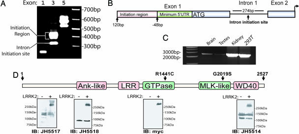
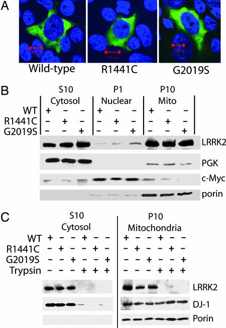
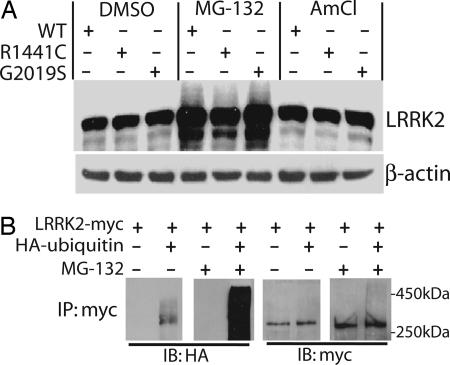
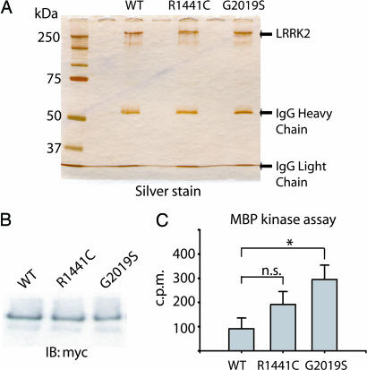
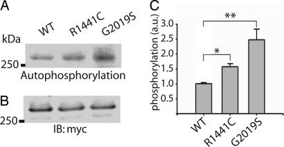
Mutations in the leucine-rich repeat kinase 2 gene (LRRK2) cause late-onset Parkinson's disease (PD) with a clinical appearance indistinguishable from idiopathic PD. Initial studies suggest that LRRK2 mutations are the most common yet identified determinant of PD susceptibility, transmitted in an autosomal-dominant mode of inheritance. Herein, we characterize the LRRK2 gene and transcript in human brain and subclone the predominant ORF. Exogenously expressed LRRK2 protein migrates at approximately 280 kDa and is present largely in the cytoplasm but also associates with the mitochondrial outer membrane. Familial-linked mutations G2019S or R1441C do not have an obvious effect on protein steady-state levels, turnover, or localization. However, in vitro kinase assays using full-length recombinant LRRK2 reveal an increase in activity caused by familial-linked mutations in both autophosphorylation and the phosphorylation of a generic substrate. These results suggest a gain-of-function mechanism for LRRK2-linked disease with a central role for kinase activity in the development of PD.


Figures

Fig. 1.
Fig. 1.
Characterization of expression of LRRK2. (A) 5′-RACE nested PCR using human total brain cDNA, as resolved on an ethidium bromide agarose gel. The exon position of the corresponding reverse oligonucleotide in the predicted LRRK2 transcript is given. (B) Diagram depicting the 5′ end of the LRRK2 gene. The ATG represents the presence of the first consensus Kozak sequence downstream of the initiation region and the start of the predominant LRRK2 ORF. (C) RT-PCR using oligonucleotide primers spanning the 3′ end of the predicted LRRK2 transcript. Sources of cDNA derived from human tissue or cell lines are indicated. (D) Protein domain structure of LRRK2 where the ankryin (Ank-like) repeat region, the leucine-rich repeat (LRR) domain, the GTPase domain, the MLK-like domain, and the WD40 domain positions are indicated. Lysate derived from HEK-293T cells transiently expressing myc-tagged LRRK2 (+) or empty (-) vector were analyzed by Western blotting by using LRRK2-specific antibodies with recognition sites near the N terminus (JH5517 and JH5518) or the C terminus (JH5514 and anti-myc).
Fig. 2.
Fig. 2.
Subcellular localization of WT and mutant LRRK2. (A) Representative merged confocal images of transiently transfected HEK-293T cells. DAPI stain is indicated as blue, and green represents myc-FITC antibody staining for LRRK2. (Red scale bars, 10 μm.) (B and C) Subcellular fractionation of HEK-293T cells transiently transfected with myc-tagged WT or mutant LRRK2 and analyzed by Western blotting. Enrichment of each fraction was assessed by stripping the membrane and reprobing with the indicated marker antibody. Two micrograms of protein per lane was loaded, and LRRK2 expression was evaluated by using anti-myc antibody.
Fig. 3.
Fig. 3.
Metabolism of WT and mutant LRRK2. (A) SH-SY5Y cells overexpressing WT or mutant LRRK2 were treated with either 5 μM MG-132 or 50 mM ammonium chloride for 24 h, and lysates were evaluated by Western blotting. LRRK2 was probed with anti-myc antibody, and the membrane was stripped and reprobed for β-actin. (B) SH-SY5Y cells were cotransfected with myc-LRRK2 with or without HA-ubiquitin, treated with or without 5 μM MG-132 for 24 h, and subjected to immunoprecipitation with anti-c-myc antibody. Immunprecipitates were probed with anti-HA or anti-c-myc antibodies as indicated.
Fig. 4.
Fig. 4.
In vitro LRRK2 kinase assay. (A) Immunoprecipitated LRRK2 preparation derived from transfected HEK-293T cells as analyzed by SDS/PAGE and subsequent silver stain. (B) Assessment of WT or mutant LRRK2 protein levels by Western blot using anti-c-myc antibody. (C) In vitro kinase activity of recombinant WT or mutant LRRK2 against biotinylated MBP. Incorporated 32P in MBP was assessed by liquid scintillation, and results are representative of at least three independent experiments. Error bars represent mean ± SEM. n.s., not significant. *, P < 0.05 assessed by two-tailed unpaired Student's t test.
Fig. 5.
Fig. 5.
Autophosphorylation of LRRK2. (A) Autoradiogram of incorporated 32P in mutant or WT LRRK2 recombinant protein resolved by SDS/PAGE. (B) LRRK2 protein levels were determined by Western blot analysis by using anti-c-myc antibody. (C) Normalization of incorporated 32P compared with LRRK2 protein content. Data represent at least three independent experiments. a.u., arbitrary units. Error bars are mean ± SEM. *, P < 0.01; **, P < 0.001 assessed by two-tailed unpaired Student's t test.
Fig. 6.
Fig. 6.
Hypothetic effect of LRRK2 G2019S mutation on kinase activity. (A) Protein alignment of the activation segment of known protein kinase. Red amino acids represent alterations in the bRaf protein that overactivate corresponding kinase activity and predispose to cancer. Green amino acids represent PD-linked mutations in LRRK2. Amino acids in bold represent the conserved protein sequence that defines the activation segment. (B) An * indicates the position of the G2019S mutation within the activation segment. The diagram is adapted from ref. .

Comment in

Similar articles

Cited by

References

    1. Funayama, M., Hasegawa, K., Kowa, H., Saito, M., Tsuji, S. & Obata, F. (2002) Ann. Neurol. 51, 296-301. - PubMed
    1. Zimprich, A., Biskup, S., Leitner, P., Lichtner, P., Farrer, M., Lincoln, S., Kachergus, J., Hulihan, M., Uitti, R. J., Calne, D. B., et al. (2004) Neuron 44, 601-607. - PubMed
    1. Paisan-Ruiz, C., Jain, S., Evans, E. W., Gilks, W. P., Simon, J., van der Brug, M., Lopez de Munain, A., Aparicio, S., Gil, A. M., Khan, N., et al. (2004) Neuron 44, 595-600. - PubMed
    1. Nichols, W. C., Pankratz, N., Hernandez, D., Paisan-Ruiz, C., Jain, S., Halter, C. A., Michaels, V. E., Reed, T., Rudolph, A., Shults, C. W., et al. (2005) Lancet 365, 410-412. - PubMed
    1. Di Fonzo, A., Rohe, C. F., Ferreira, J., Chien, H. F., Vacca, L., Stocchi, F., Guedes, L., Fabrizio, E., Manfredi, M., Vanacore, N., et al. (2005) Lancet 365, 412-415. - PubMed

Publication types

MeSH terms

Substances