. 2005 Nov;71(11):6845-55.
doi: 10.1128/AEM.71.11.6845-6855.2005.

Identification and characterization of a novel biotin biosynthesis gene in Saccharomyces cerevisiae

Affiliations

Identification and characterization of a novel biotin biosynthesis gene in Saccharomyces cerevisiae

Hong Wu et al. Appl Environ Microbiol. 2005 Nov.

Abstract

Yeast Saccharomyces cerevisiae cells generally cannot synthesize biotin, a vitamin required for many carboxylation reactions. Although sake yeasts, which are used for Japanese sake brewing, are classified as S. cerevisiae, they do not require biotin for their growth. In this study, we identified a novel open reading frame (ORF) in the genome of one strain of sake yeast that we speculated to be involved in biotin synthesis. Homologs of this gene are widely distributed in the genomes of sake yeasts. However, they are not found in many laboratory strains and strains used for wine making and beer brewing. This ORF was named BIO6 because it has 52% identity with BIO3, a biotin biosynthesis gene of a laboratory strain. Further research showed that yeasts without the BIO6 gene are auxotrophic for biotin, whereas yeasts holding the BIO6 gene are prototrophic for biotin. The BIO6 gene was disrupted in strain A364A, which is a laboratory strain with one copy of the BIO6 gene. Although strain A364A is prototrophic for biotin, a BIO6 disrupted mutant was found to be auxotrophic for biotin. The BIO6 disruptant was able to grow in biotin-deficient medium supplemented with 7-keto-8-amino-pelargonic acid (KAPA), while the bio3 disruptant was not able to grow in this medium. These results suggest that Bio6p acts in an unknown step of biotin synthesis before KAPA synthesis. Furthermore, we demonstrated that expression of the BIO6 gene, like that of other biotin synthesis genes, was upregulated by depletion of biotin. We conclude that the BIO6 gene is a novel biotin biosynthesis gene of S. cerevisiae.


Figures
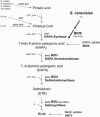
FIG. 1.
FIG. 1.
Pathways of biotin biosynthesis.
FIG. 2.
FIG. 2.
Cloning of the BIO6 gene of K7. (A) Structure of contig 4.1. The arrows indicate protein coding regions and orientations. E, EcoRI; Ev, EcoRV; H, HindIII; N, NotI; P, PstI; S, SalI; Sp, SphI; X, XbaI. The COS-55-F fragment is indicated by a bar. (B) PFGE Southern blot analyses of S288C (lanes 1 and 3) and K7 (lanes 2 and 4). Chromosome DNAs prepared from approximately the same amount of cells were used for PFGE Southern blot analysis using COS-55-F (lanes 1 and 2) and COS-55-R (lanes 3 and 4) fragments as probes. The presumed chromosome numbers are indicated.
FIG. 3.
FIG. 3.
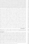
Nucleotide and deduced amino acid sequences of the BIO6 gene. The COS-55-F sequence is indicated by underlining. The SAM and PLP binding sites are indicated by double underlining.
FIG. 4.
FIG. 4.
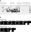
Relationship between the BIO6 gene and biotin prototrophy. (A) PFGE Southern blot analysis of the BIO6 gene in various yeast strains. Chromosome DNAs prepared from approximately the same amount of cells were used for PFGE Southern blot analysis using the COS-55-F fragment as a probe. (B) Biotin auxotrophic assay. Yeast strains with BIO6 (top) or without BIO6 (bottom) were streaked on SM+Biotin and SM-Biotin and incubated at 30°C for 6 days. Strain X2180 (top) was used as a negative control.
FIG. 5.
FIG. 5.
Cloning and disruption of the BIO6 gene in A364A. (A) PFGE Southern blot analyses of BIO6 of K7 and A364A. Chromosome DNAs prepared from approximately the same amount of cells were used for PFGE Southern blot analysis using the COS-55-F fragment as a probe. The presumed chromosome numbers are indicated. (B) Gene disruption of the BIO6 gene in A364A. A 1.5-kb fragment containing the BIO6 gene of A364A was amplified and cloned. The Geneticin resistance gene kanMX4, which was used as a selectable marker, was inserted in this fragment. Strain A364A was then transformed with part of this fragment. (C) Biotin auxotrophic assay. A364A, YHW11 (bio6Δ), YHW13 harboring an empty vector in YHW11 (bio6Δ-101), and YHW12 harboring a vector containing BIO6 of A364A (bio6Δ-BIO6) were tested. Cells of these strains were streaked on SM+Biotin and SM-Biotin and incubated at 30°C for 7 days.
FIG. 6.
FIG. 6.
Effects of biotin synthesis intermediates on BIO6- or BIO3-disrupted strains. A364A, YHW11 (bio6Δ), and YHW14 (bio3Δ) were streaked on SM-Biotin, SM+KAPA, SM+DAPA, and SM+DTB and incubated at 30°C for 7 days.
FIG. 7.
FIG. 7.
Effect of biotin on BIO6 gene expression. Each lane was loaded with 5 μg total RNA from of K7 or A364A cells grown in the presence and absence of biotin. A COS-55-F fragment was used as a probe. Lanes 1 and 2, K7 cells grown in SM+Biotin and SM-Biotin, respectively; lanes 3 and 4, A364A cells grown in SM+Biotin and SM-Biotin, respectively. The ACT1 signal and ethidium bromide staining were determined as loading controls.
FIG. 8.
FIG. 8.
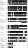
Sequence comparison of K7 Bio6p and E. coli bioA and bioF products. Conserved amino acids are indicated by white letters on black backgrounds. The SAM and PLP binding sites are indicated by asterisks and dots, respectively.
FIG. 9.
FIG. 9.
Multiple sequence alignment of Bio6p. Amino acid sequence alignment of Bio6p of K7 with those of A364A, S. bayanus (accession no. AACA0100015), S. kudriavzevii (accession no. AACI01000492), S. paradoxus (accession no. AABY01000445 and AABY01000593) and S. mikatae (accession no. AABZ01000331). Amino acids that are conserved among at least five of the six strains are indicated by white letters on black backgrounds. Positions of the SAM and PLP binding sites are shown by asterisks and dots, respectively.

Similar articles

Cited by

References

    1. Adams, A., D. E. Gottschling, C. Kaiser, and T. Stearns. 1997. Methods in yeast genetics. Cold Spring Harbor Laboratory Press, Cold Spring Harbor, N.Y.
    1. Baldet, P., and M. L. Ruffet. 1996. Biotin synthesis in higher plants: isolation of a cDNA encoding Arabidopsis thaliana bioB-gene product equivalent by functional complementation of a biotin auxotroph mutant bioB105 of Escherichia coli K12. C. R. Acad. Sci. Ser. III 319:99-106. - PubMed
    1. Becker, D. M., and L. Guarente. 1991. High-efficiency transformation of yeast by electroporation. Methods Enzymol. 194:182-187. - PubMed
    1. Betz, H., H. Hinze, and H. Holzer. 1974. Isolation and properties of two inhibitors of proteinase B from yeast. J. Biol. Chem. 249:4515-4521. - PubMed
    1. Bower, S., J. B. Perkins, R. R. Yocum, C. L. Howitt, P. Rahaim, and J. Pero. 1996. Cloning, sequencing, and characterization of the Bacillus subtilis biotin biosynthetic operon. J. Bacteriol. 178:4122-4130. - PMC - PubMed

Publication types

MeSH terms

Associated data