. 2005 Nov;71(11):7334-44.
doi: 10.1128/AEM.71.11.7334-7344.2005.

Antagonists of Hsp16.3, a low-molecular-weight mycobacterial chaperone and virulence factor, derived from phage-displayed peptide libraries

Affiliations

Antagonists of Hsp16.3, a low-molecular-weight mycobacterial chaperone and virulence factor, derived from phage-displayed peptide libraries

Abhik Saha et al. Appl Environ Microbiol. 2005 Nov.

Abstract

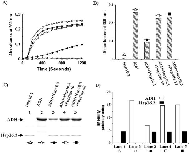
The persistence of Mycobacterium tuberculosis is a major cause of concern in tuberculosis (TB) therapy. In the persistent mode the pathogen can resist drug therapy, allowing the possibility of reactivation of the disease. Several protein factors have been identified that contribute to persistence, one of them being the 16-kDa low-molecular-weight mycobacterial heat shock protein Hsp16.3, a homologue of the mammalian eye lens protein alpha-crystallin. It is believed that Hsp16.3 plays a key role in the persistence phase by protecting essential proteins from being irreversibly denatured. Because of the close association of Hsp16.3 with persistence, an attempt has been made to develop inhibitors against it. Random peptide libraries displayed on bacteriophage M13 were screened for Hsp16.3 binding. Two phage clones were identified that bind to the Hsp16.3 protein. The corresponding synthetic peptides, an 11-mer and a 16-mer, were able to bind Hsp16.3 and inhibit its chaperone activity in vitro in a dose-dependent manner. Little or no effect of these peptides was observed on alphaB-crystallin, a homologous protein that is a key component of human eye lens, indicating that there is an element of specificity in the observed inhibition. Two histidine residues appear to be common to the selected peptides. Nuclear magnetic resonance studies performed with the 11-mer peptide indicate that in this case these two histidines may be the crucial binding determinants. The peptide inhibitors of Hsp16.3 thus obtained could serve as the basis for developing potent drugs against persistent TB.


Figures

FIG. 1.
FIG. 1.
Reverse phage ELISA to confirm the binding of phage clones displaying peptide sequences, Seq ID 9 and 21, to Hsp16.3. The phage clones were amplified, concentrated by PEG precipitation, and reacted with either Hsp16.3 or nonspecific target BSA coated on the wells of a microtiter plate. Unbound phage was removed by washing with TBST (0.5% Tween 20), and bound phage was detected with horseradish peroxidase-conjugated anti-M13 monoclonal antibody (1:5,000) and ABTS substrate. Color development was monitored spectrophotometrically at 405 nm. Each experiment was done in triplicate. The bar graph shows binding activity of phage clones displaying Seq ID 9 (□) and Seq ID 21 (▪) to Hsp16.3 and BSA (background binding). The error bars represent the SE.
FIG. 2.
FIG. 2.
Determination of the dissociation constant and stoichiometry of peptide binding to Hsp16.3. Anisotropy increase of FITC-labeled peptide 10 (A) and peptide 22 (B) as a function of Hsp16.3 concentration in 50 mM sodium phosphate buffer (pH 7.5) containing 300 mM NaCl at room temperature. Anisotropy values are means ± the SE (indicated by error bars) of three independent experiments. The excitation and emission wavelengths were at 495 and 519 nm, respectively. In case of peptide 10, stoichiometry was determined (C) from the dose-dependent quenching of the intrinsic tyrosine fluorescence of Hsp16.3 in the same buffer conditions. The lines were drawn to determine binding stoichiometry.
FIG. 3.
FIG. 3.
Dose-dependent inhibition of chaperone activity of Hsp16.3 by peptides 10 and 22. Aggregation assays were performed by heating 5 μM ADH at 50°C directly in a spectrophotometer using a thermostated cuvette holder in a total reaction volume of 500 μl in 100 mM sodium phosphate buffer (pH 7.0) containing 100 mM NaCl. The absorbance was monitored at 360 nm for 1,200 s, and readings were taken at every 100-s interval. ADH aggregates upon heating (A, B, D, and E; ▴). Incorporation of Hsp16.3 at a molar ratio of 5:4 (A and D) or alphaB-crystallin at a molar ratio of 1:1 (B and E) results in a decline in aggregation activity (•). Then increasing concentrations of Hsp16.3 binding peptide 10 (A and B) and peptide 22 (D and E) were added as indicated at the following concentrations: 25 μM (□), 50 μM (▪), and 100 μM (▵). Stars represent the ADH aggregation in the presence of peptide 10 (A and B) and peptide 22 (D and E). The IC50 for peptide 10 (C) and peptide 22 (F) was calculated from the plot of the percent residual chaperone activity against the peptide concentration. Each datum point in panels C and F is an average of three independently derived results. Error bars indicate the SE.
FIG. 4.
FIG. 4.
Effect of peptides on the stability of the Hsp16.3-MDH complex. (A) MDH (2 μM) either alone or after being mixed with Hsp16.3 (80 μM) was heated to 50°C for 30 min, followed by centrifugation at 15,000 × g. The supernatants and pellets were analyzed by SDS-PAGE on a 15% gel. Lanes marked with black dots represent pellet fractions in all cases. The MDH-Hsp16.3 complex was then formed in the presence of inhibitory peptide 10 or peptide 22 (B and C), respectively. The molar excesses of the peptides used relative to Hsp16.3 are indicated below the lanes. The bands were scanned densitometrically, and the fraction of the protein (either Hsp16.3 or MDH) that appeared in the supernatant was plotted as the percent solubility against the molar excess of peptide 10 or peptide 22 (D and E), respectively. (F and G) The effect of peptides 10 and 22 on the solubility of Hsp16.3 was tested using the indicated molar excess. (H) The comparative effect of peptide 22 (lanes marked with black dots) on alphaB-crystallin was determined side by side with the Hsp16.3 control. (I) Similarly, the effect of peptide 22 on the formation of a soluble complex between alphaB-crystallin and MDH was tested alongside an Hsp16.3-MDH control. In the last two experiments, a peptide molar excess of 15 over either Hsp16.3 or alphaB-crystallin was used.
FIG. 5.
FIG. 5.
Peptide-mediated inactivation of Hsp16.3 under conditions used for spectrophotometric assay of chaperone activity. (A) The chaperone activity was measured spectrophotometrically as described in Fig. 3. Aggregation of ADH (5 μM) without chaperone is represented by the curve marked by white circles, protection by Hsp16.3 (4 μM) by black circles, and inhibition of Hsp16.3 by peptides 10 and 22 (200 μM) by white and black squares, respectively. Aggregation of Hsp16.3 alone is indicated by white triangles. (B) The steady-state aggregations (1,200 s) observed in panel A are graphically represented. (C) The aggregated samples were centrifuged, and the pellet fractions were analyzed by SDS-PAGE on a 15% gel; lanes corresponding to the aggregation curves are indicated by identical symbols. The combinations of proteins and peptides used are indicated on the top. The bar diagram (D) shows the band intensities of the ADH (□) and Hsp16.3 (▪) components in each lane.
FIG. 6.
FIG. 6.
Changes in chemical shift of peptide 10 residues due to interaction with Hsp16.3. (A) Overlay 2D TOCSY spectra in the fingerprint (selected) region of 2 mM peptide 10 in 20 mM sodium phosphate buffer (pH 7.0) containing 250 mM NaCl in 90% H2O and 10% D2O at 5°C in the absence (red) or in the presence (blue) of 0.1 mM Hsp16.3 protein. Amino acids are designated by using the one-letter code. (B) Graphical representation of chemical shift differences (Δ δ Hz) of amide protons of peptide 10 residues in buffer solution in the presence or absence of Hsp16.3, as mentioned in the legend to panel A.

References

    1. Andreu, D., and L. Rivas. 1998. Animal antimicrobial peptides: an overview. Biopolymers 47:415-433. - PubMed
    1. Appeldoorn, C. C., T. J. Molenaar, A. Bonnefoy, S. H. van Leeuwen, P. A. Vandervoort, M. F. Hoylaerts, T. J. van Berkel, and E. A. Biessen. 2003. Rational optimization of a short human P-selectin-binding peptide leads to nanomolar affinity antagonists. J. Biol. Chem. 278:10201-10207. - PubMed
    1. Barker, W. C., J. S. Garavelli, H. Huang, P. B. McGarvey, B. C. Orcutt, G. Y. Srinivasarao, C. Xiao, L. L. Yeh, R. S. Ledley, J. F. Janda, F. Pfeiffer, H. Mewes, A. Tsugita, and C. Wu. 2000. The protein information resource (PIR). Nucleic Acids Res. 28:41-44. - PMC - PubMed
    1. Barry, C., III. November2002. Method of attenuating pathogenic mycobacteria and strains of mycobacteria so attenuated. U.S. patent 6,403,100.
    1. Bhattacharyya, T., A. Bhattacharyya, and S. Roy. 1991. A fluorescence spectroscopic study of glutaminyl-tRNA synthetase from Escherichia coli and its implications for the enzyme mechanism. Eur. J. Biochem. 200:739-745. - PubMed

Publication types

MeSH terms

LinkOut - more resources