. 2005 Nov 16;24(22):3963-73.
doi: 10.1038/sj.emboj.7600857. Epub 2005 Nov 3.

Concomitant loss of proapoptotic BH3-only Bcl-2 antagonists Bik and Bim arrests spermatogenesis

Affiliations

Concomitant loss of proapoptotic BH3-only Bcl-2 antagonists Bik and Bim arrests spermatogenesis

Leigh Coultas et al. EMBO J. .

Abstract

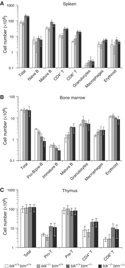
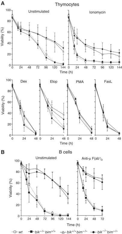
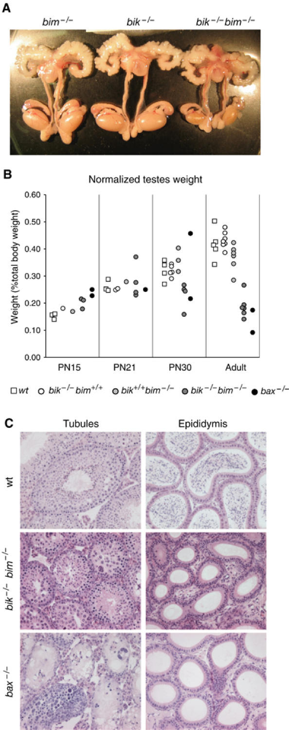
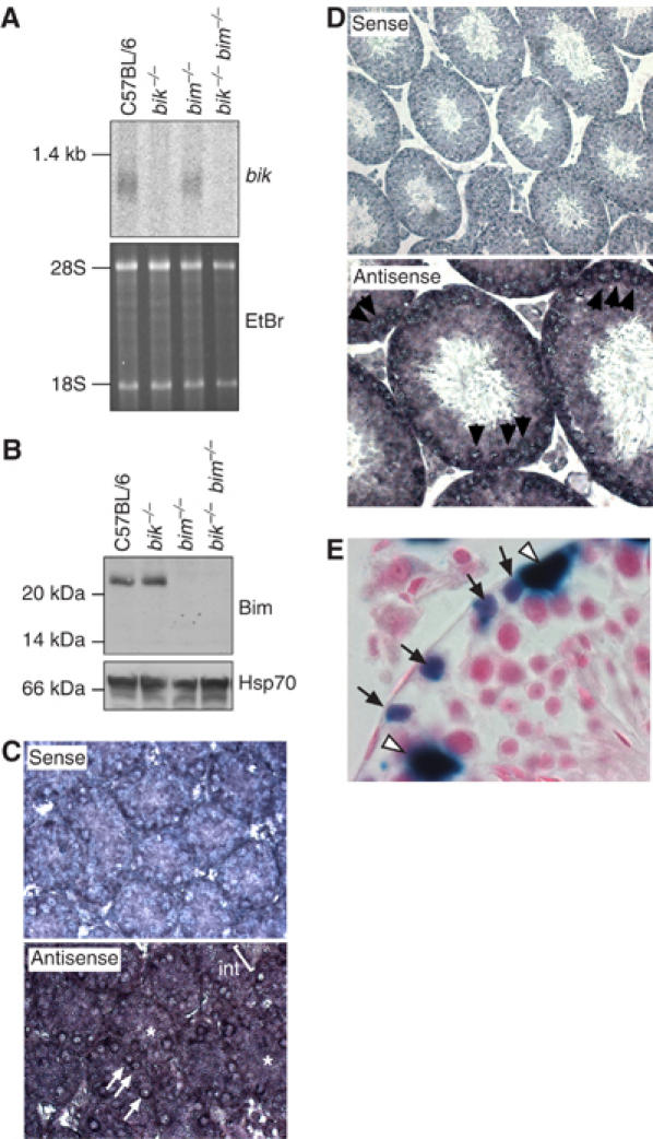
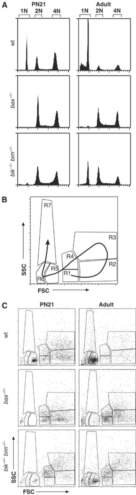
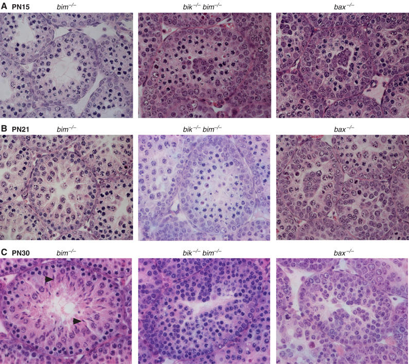
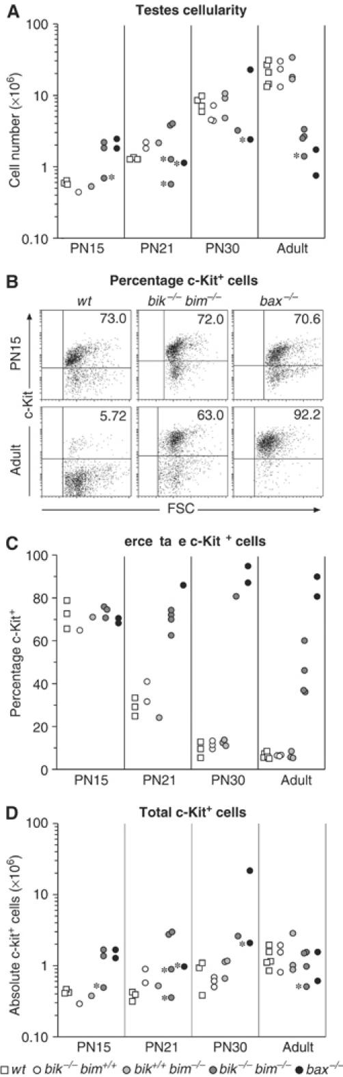
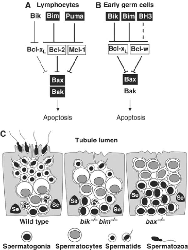
The BH3-only proteins of the Bcl-2 family initiate apoptosis through the activation of Bax-like relatives. Loss of individual BH3-only proteins can lead either to no phenotype, as in mice lacking Bik, or to marked cell excess, as in the hematopoietic compartment of animals lacking Bim. To investigate whether functional redundancy with Bim might obscure a significant role for Bik, we generated mice lacking both genes. The hematopoietic compartments of bik-/-bim-/- and bim-/- mice were indistinguishable. However, although testes develop normally in mice lacking either Bik or Bim, adult bik-/-bim-/- males were infertile, with reduced testicular cellularity and no spermatozoa. The testis of young bik-/-bim-/- males, like those lacking Bax, exhibited increased numbers of spermatogonia and spermatocytes, although loss of Bik plus Bim blocked spermatogenesis somewhat later than Bax deficiency. The initial excess of early germ cells suggests that spermatogenesis fails because supporting Sertoli cells are overwhelmed. Thus, Bik and Bim share, upstream of Bax, the role of eliminating supernumerary germ cells during the first wave of spermatogenesis, a process vital for normal testicular development.


Figures

Figure 1
Figure 1
Cell type composition of hematopoietic organs from wt (bik+/+bim+/+), bik−/−bim+/+, bik+/+bim−/− and bik−/−bim−/− mice. (A) The total number of splenocytes from 8- to 13-week-old wt, bik−/−bim+/+, bik+/+bim−/− and bik−/−bim−/− mice was determined and the number of naïve (sIgM+ sIgD) and mature B cells (sIgMlo sIgDhi), mature T cells (CD4+ CD8 or CD4 CD8+), macrophages (Mac1+ Gr-l), granulocytes (Mac1+ Gr-l+) and nucleated erythroid progenitors (Ter119+) was determined by FACS analysis. (B) The total number of bone marrow cells from 8- to 13-week-old wt, bik−/−bim+/+, bik+/+bim−/− and bik−/−bim−/− mice was determined and the number of pro-B and pre-B cells (B220+ sIgM sIgD), immature (sIgM+ sIgD) and mature (sIgMlo sIgDhi) B cells, macrophages (Mac1 Grl+) and granulocytes (Mac1+ Grl+) was determined by FACS analysis. (C) The total number of thymocytes from 8- to 13-week-old wt, bik−/−bim+/+, bik+/+bim−/− and bik−/−bim−/− mice was determined and the number of pro-T (CD48), pre-T (CD4+ 8+) and mature T (CD4+ 8 or CD48+) cells was determined by FACS analysis. Data shown represent means±s.d. of 3–5 mice of each genotype.
Figure 2
Figure 2
Susceptibility of bik−/−bim−/− lymphocytes to apoptotic stimuli. (A) Thymocytes from wt (bik+/+bim+/+), bik−/−bim+/+, bik+/+bim−/− and bik−/−bim−/− mice were harvested and cultured in the presence of dexamethasone (Dex 10−6 M), etoposide (Etop 1 μg/ml), PMA (2 ng/ml), ionomycin (1 μg/ml), crosslinked FasL (100 ng/ml) or in the absence of exogenous stimuli (unstimulated) for the indicated times. (B) Mature B cells were negatively sorted from lymph nodes of wt (bik+/+bim+/+), bik−/− bim+/+, bik+/+ bim−/− and bik−/−bim−/− mice and cultured in the presence of anti-IgM crosslinking F(ab′) antibody fragments at 5 μg/ml or in the absence of exogenous stimuli (unstimulated) for the indicated times. Data shown represent means±s.d. of 4–6 mice of each genotype.
Figure 3
Figure 3
Reduced testicular size and absence of spermatozoa in bik−/−bim−/− males. (A) Dissected reproductive tracts from representative bik−/−bim+/+, bik+/+bim−/− and bik−/−bim−/− males showing reduced testicular size, but otherwise normal appearance. Testes of bik−/−bim+/+, bik+/+bim−/− mice were indistinguishable from those of wt males. (B) Weights of testes from wt, bik−/−bim+/+, bik+/+bim−/−, bik−/−bim−/− and bax−/− males at PN day 15, 21, 30 and adult. Each data point represents the weight of one testis from a single male normalized to body weight. (C) Histological appearance of tubules and epididymis from testes of adult wt, bik−/−bim−/− and bax−/− males.
Figure 4
Figure 4
Expression of Bik and Bim in testes. (A) Northern blot analysis of poly(A)+ RNA extracted from the testes of adult wt C57BL/6, bik−/−, bim−/− and bik−/−bim−/− mice. The blot was probed with a cDNA probe containing the entire bik coding region. Ethidium bromide stain of the RNA is shown as a loading control; 28S and 18S rRNA species are indicated. (B) Western blot analysis of Bim protein expression in whole-cell lysates extracted from the testes of adult wt C57BL/6, bik−/−, bim−/− and bik−/−bim−/− mice. Probing with an anti-Hsp70 mAb was used as a loading control. (C) Bik gene expression assessed in PN5 testes by in situ hybridization, showing a strong signal in spermatogonia (arrows) and a less intense signal in the Sertoli cell cytoplasm (asterisk), while the interstitial signal was weak to absent (Int). Sense probe (top panel) is included as a negative control. (D) Bik gene expression in the adult testis was assessed by in situ hybridization, showing strongest signal with spermatocytes (arrows). Sense probe (top panel) is included as a negative control. (E) β-Galactosidase assay as a surrogate for bik expression in bik+/− adult testes. β-Galactosidase activity was evident in spermatogonia (arrows) and Sertoli cells (open arrow head).
Figure 5
Figure 5
Profiling germ cell development in bik−/−bim−/− mice by FACS. (A) DNA content of bik−/−bim−/− male germ cells was assessed by PI staining and FACS analysis and compared to that in wt and bax−/− germ cells. Cells with sub-1N DNA content represent spermatozoa with condensed chromatin, such that PI cannot fully access the DNA. (B) Schematic diagram of germ cell analysis by FACS. Development of mature spermatozoa from spermatogonia (R1 and R4) follows the direction of the arrow on the FSC/SSC plot. The premeiotic 2N spermatogonia appear in regions R1 and R4. On replicating their DNA to become 4N primary spermatocytes, they shift to R2. When the spermatocytes condense their chromosomes, they shift into R3 as late-stage spermatocytes. The first meiotic division produces 2° spermatocytes, which transit back through R1 and R4 before entering R5 as round spermatids after the second meiotic division. As spermatids elongate and condense their DNA they occupy R5 and R6, and finally on becoming mature spermatozoa they reach R7 (although mature spermatozoa contribute to R5, R6 and R7) (Malkov et al, 1998). (C) FACS profiles of germ cell suspensions prepared from wt, bik−/−bim−/− and bax−/− testes at PN21 and adults, showing arrested germ cell development at the early primary spermatocyte stage in bax−/− mice and at the round spermatid stage in bik−/−bim−/− mice.
Figure 6
Figure 6
Histological appearance of developing bik−/−bim−/− testes. At all ages, the bim−/− testis (left panels) was indistinguishable from a wt or bik−/− testis (data not shown). (A) Sections of PN15 testes, stained with hematoxylin and eosin, showing increased thickness of the spermatogonial (outer) cell layer and cell clusters in the centre of bik−/−bim−/− and bax−/− tubules compared to controls. (B) Sections of PN21 testes, stained with hematoxylin and eosin, showing that bik−/−bim−/− testes have increased layers of spermatogonia, compared to bim−/− testes. (C) Sections of PN30 testes, stained with hematoxylin and eosin, showing accumulation of spermatocytes in bik−/−bim−/− testes. By comparison, bax−/− tubules show preferential accumulation of spermatogonia and some early spermatocytes. Some bik−/−bim−/− tubules contained round spermatids, but not the elongating spermatids (arrowheads) observed in bim−/−, bik−/− and wt controls.
Figure 7
Figure 7
Accumulation of c-Kit+ germ cells in bik−/−bim−/− testes. (A) Total cellularity of wt, bik−/−, bim−/−, bik−/−bim−/− and bax−/− testes taken at PN15, PN21, PN30 and adult (8–15 weeks old). Each data point represents the number of germ cells isolated from a single testis of one animal. Asterisks mark testes taken from runts. (B) Representative FACS profiles of PN15 and adult (8–15 weeks) wt, bik−/−bim−/− and bax−/− testes stained for c-Kit expression. (C) The percentage of c-Kit+ cells in germ cell preparations from PN15, PN21, PN30 and adult (8–15 weeks) wt, bik−/−, bim−/−, bik−/−bim−/− and bax−/− testes. Each data point represents the percentage of c-Kit+ germ cells from a single testis of one animal. (D) Absolute numbers of c-Kit+ cells in the germ cell preparations of PN15, PN21, PN30 and adult testes from wt, bik−/−, bim−/−, bik−/−bim−/− and bax−/− testes. Each data point represents the number of germ cells isolated from a single testis of one animal. Asterisks mark testes taken from runts.
Figure 8
Figure 8
Model for the disrupted spermatogenesis in bik−/−bim−/− mice. The key regulators of apoptosis within lymphocytes (A) and early germ cells (B) are compared (see text). For each cell type, boxed genes indicate dominant Bcl-2 family regulators of apoptosis and thick lines denote dominant pathways. The BH3-only protein postulated to cooperate with Bim and Bik in germ cells is indicated as BH3. Bcl-xL is assumed to have a greater role in spermatogonia than Bcl-w because loss of a single allele of the former but not the latter impairs male germ cell apoptosis (Kasai et al, 2003). (C) During the first wave of normal spermatogenesis (wt), the excess of germ cells, particularly spermatogonia, is eliminated by apoptosis (crinkly cell) because the Sertoli cells (Se) cannot provide sufficient support. In the bik−/−bim−/− and bax−/− testes, the failure of apoptosis during the first wave produces an abnormal excess of spermatogonia and spermatocytes that is presumed to overwhelm the Sertoli cells, leading to aborted testicular development.

References

    1. Adams JM (2003) Ways of dying: multiple pathways to apoptosis. Genes Dev 17: 2481–2495 - PubMed
    1. Akiyama T, Bouillet P, Miyazaki T, Kadono Y, Chikuda H, Chung UI, Fukuda A, Hikita A, Seto H, Okada T, Inaba T, Sanjay A, Baron R, Kawaguchi H, Oda H, Nakamura K, Strasser A, Tanaka S (2003) Regulation of osteoclast apoptosis by ubiquitylation of proapoptotic BH3-only Bcl-2 family member Bim. EMBO J 22: 6653–6664 - PMC - PubMed
    1. Allan DJ, Harmon BV, Kerr JFR (1987) Cell death in spermatogenesis. In Perspectives on Mammalian Cell Death, Potten CS (ed) pp 229–258. Oxford: Oxford University Press
    1. Bouillet P, Cory S, Zhang L-C, Strasser A, Adams JM (2001) Degenerative disorders caused by Bcl-2 deficiency are prevented by loss of its BH3-only antagonist Bim. Dev Cell 1: 645–653 - PubMed
    1. Bouillet P, Metcalf D, Huang DCS, Tarlinton DM, Kay TWH, Köntgen F, Adams JM, Strasser A (1999) Proapoptotic Bcl-2 relative Bim required for certain apoptotic responses, leukocyte homeostasis, and to preclude autoimmunity. Science 286: 1735–1738 - PubMed

Publication types

MeSH terms