. 2005 Nov 16;24(22):3834-45.
doi: 10.1038/sj.emboj.7600847. Epub 2005 Nov 3.

PKCepsilon-mediated phosphorylation of vimentin controls integrin recycling and motility

Affiliations

PKCepsilon-mediated phosphorylation of vimentin controls integrin recycling and motility

Johanna Ivaska et al. EMBO J. .

Abstract

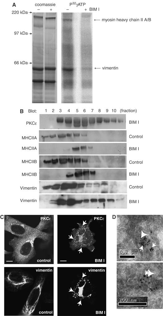
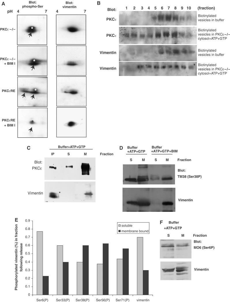
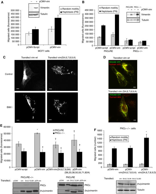
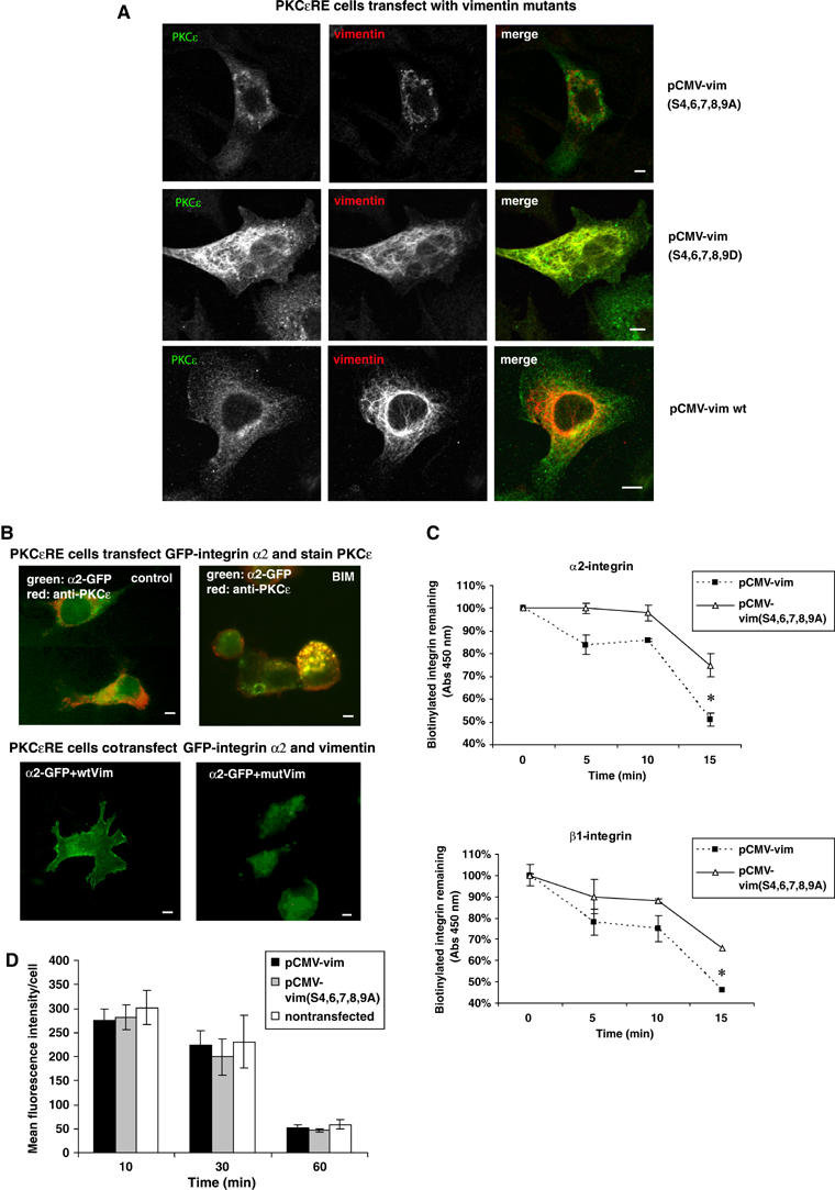
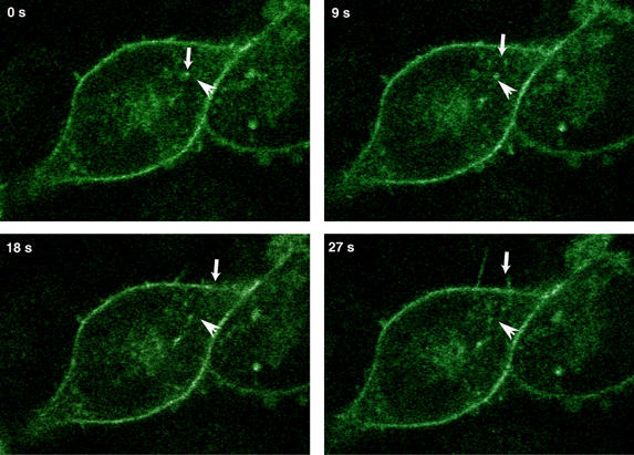
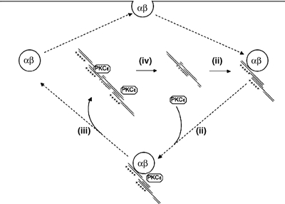
PKCepsilon controls the transport of endocytosed beta1-integrins to the plasma membrane regulating directional cell motility. Vimentin, an intermediate filament protein upregulated upon epithelial cell transformation, is shown here to be a proximal PKCepsilon target within the recycling integrin compartment. On inhibition of PKC and vimentin phosphorylation, integrins become trapped in vesicles and directional cell motility towards matrix is severely attenuated. In vitro reconstitution assays showed that PKCepsilon dissociates from integrin containing endocytic vesicles in a selectively phosphorylated vimentin containing complex. Mutagenesis of PKC (controlled) sites on vimentin and ectopic expression of the variant leads to the accumulation of intracellular PKCepsilon/integrin positive vesicles. Finally, introduction of ectopic wild-type vimentin is shown to promote cell motility in a PKCepsilon-dependent manner; alanine substitutions in PKC (controlled) sites on vimentin abolishes the ability of vimentin to induce cell migration, whereas the substitution of these sites with acidic residues enables vimentin to rescue motility of PKCepsilon null cells. Our results indicate that PKC-mediated phosphorylation of vimentin is a key process in integrin traffic through the cell.


Figures

Figure 1
Figure 1
PKC substrates on endocytic integrin-containing vesicles. (A) To identify proximal PKC targets in the vesicular compartment, biotinylated vesicles derived from BIM-I treated PKCɛ expressing cells (PKCɛRE) were resuspended in a small volume of buffer and incubated in cytosol derived from PKCɛ−/− cells in the presence of calyculin A, P32γATP and PKC inhibitor BIM-I where indicated for 1 h at 37°C. Following the reaction, all biotinylated proteins were collected on streptavidin-Dynabeads and resolved on SDS–PAGE followed by Coomassie staining and autoradiography. BIM-I sensitive phosphorylated bands were cut from the gel, trypsinised and identified as vimentin and myosin heavy chain II A or B using MS fingerprinting. (B) The subcellular localisation of vimentin and MHCII alter upon PKC inhibition. PKCɛRE cells were treated for 90 min with BIM-I or left untreated, followed by sucrose gradient fractionation (see Materials and methods). The proteins in the fractions were recovered by TCA precipitation and subjected to Western blot analysis with anti- PKCɛ, anti-vimentin, anti-MHCIIA and anti-MHCIIB antibodies. Upon BIM-I treatment, vimentin was found to accumulate in the PKCɛ positive dense compartment (fractions 7–9). (C) BIM-I treatment induces vimentin fragmentation and the formation of PKCɛ positive vesicles. Dual-colour immunofluorescence staining of vimentin and PKCɛ in PKCɛRE cells is shown. The cells were plated and allowed to spread of fibronectin for 30 min, following 90 min incubation either untreated (control) or 1 μM BIM-I (BIM-I). Arrows indicate some of the areas of overlap in the PKCɛ and vimentin stainings in BIM-I treated cells. Bar 10 μm. (D) Enrichment of vimentin and PKCɛ in vesicles isolated from BIM-I treated PKCɛRE cells. The immunoelectron micrographs show double-labelling of PKCɛ (10 nm protein A-gold, big arrowheads) and vimentin (15 nm protein A-gold, small arrows).
Figure 2
Figure 2
PKCɛ-dependent phosphorylation of vimentin and regulation of vimentin association in the vesicular compartment. (A) PKCɛ null (−/−) and PKCɛRE cells were treated with PKC inhibitor BIM-I where indicated and whole-cell lysates of equal protein loading were resolved with 2D gel electrophoresis and transferred onto nitrocellulose. Serine-phosphorylation was detected with blotting using an anti-phospho-serine antibody (left-hand panels) followed by stripping and reprobing with anti-vimentin to detect the localisation of the endogenous proteins (right-hand panels). In addition to vimentin, the phospho-serine antibody detects an unidentified protein spot migrating at slightly higher molecular weight than vimentin (marked ‘*'). The phospho-serine signal corresponding to vimentin is indicated with arrows. (B) Biotinylated vesicles from BIM-I treated PKCɛRE cells were resuspended in a small volume of buffer and incubated in cytosol derived from PKCɛ−/− cells in the presence of ATP and GTP, to support PKC catalytic activity, or buffer alone as indicated for 1 h at 37°, followed by refractionation on an equilibrium gradient. Biotinylated proteins from each fraction were collected on streptavidin-Dynabeads, the bound proteins were subjected to Western blot analysis following SDS–PAGE electrophoresis. (C) Isolated vesicles from BIM-I treated PKCɛRE cells were resuspended in a small volume of buffer and incubated for 1 h at 37°C in buffer in the presence of ATP and GTP. The reaction was diluted into two volumes of ice cold HEPES-buffer and sedimented at 100 000 g to recover the remaining membrane-bound proteins. Vimentin was immunoprecipitated from the soluble fraction (IP) and the remaining soluble proteins were concentrated with TCA precipitation. Proteins from all three fractions were assayed by Western blot analysis to detect PKCɛ and vimentin. (D) Isolated vesicles from BIM-I treated PKCɛRE cells were resuspended to a small volume of buffer and incubated for 1 h at 37°C in buffer in the presence of ATP and GTP and BIM I where indicated. The reaction was diluted into two volumes of ice cold HEPES-buffer and sedimented at 100 000 g to recover the remaining membrane bound proteins; the remaining soluble proteins were concentrated with TCA precipitation. Proteins from both fractions were assayed with Western blot analysis to detect vimentin phosphorylation at specific sites. Vimentin distribution was detected by stripping and reprobing after phospho-antibody detection. A representative blot with vimentin anti-phosphoserine 38 is shown in (D). (E) The histogram shows quantification of the distribution of vimentin phosphorylated at the sites indicated in the membrane bound and soluble fractions in a representative experiment. (F) A representative blot with vimentin anti-phosphoserine 6 is shown.
Figure 3
Figure 3
Vimentin induced cell motility is dependent on PKCɛ and the phosphorylation of vimentin. (A) MCF7 cells and (B) PKCɛ null (−/−) and PKCɛRE cells were transiently cotransfected with empty vector (pCMV-Script) or wild-type (pCMV-vim) human vimentin together with a luciferase encoding vector (transfection efficiency ∼80%). The bottoms of Transwell filters were coated using 10 μg/ml BSA (random motility) or FN (haptotaxis). At 36 h post-transfection, 104 cells per well were allowed to migrate for 16 h. The migrated cells were detached with trypsin, lysed and quantified using a DNA dye (see Materials and methods). An aliqout of the cells was also lysed and assayed for luciferase activity and migration was normalised to transfection efficiency. Inserted panels show vimentin expression levels before and after transient transfections with pCMV-vim in these cells. (C) Immunofluorescence staining of vimentin in PKCɛRE cells is shown. The cells were transiently transfected with wt human vimentin or vimentin(S4,6,7,8,9A). At 36 h post-transfection, the cells were plated and allowed to spread on fibronectin for 30 min, following 90 min incubation either untreated (control) or 1 μM BIM-I (BIM-I). Bar 10 μm. (D) Immunofluorescence staining of endogenous mouse vimentin (green) and ectopically expressed human vimentin (red) in PKCɛRE cells are shown. The cells were transiently transfected with wt human vimentin or vimentin(S4,6,7,8,9A). At 36 h post-transfection, the cells were plated and allowed to spread on fibronectin for 1 h. Bar 10 μm. (E) PKCɛRE cells were transiently cotransfected with empty vector (pCMV-Script), wt human vimentin (pCMV-vim), vimentin(S4,6,7,8,9A) or vimentin(S6,33,38,50,55,71,82A) together with the luciferase encoding vector. Transfected cell haptotaxis towards fibronectin was assayed as above (means±s.d., n=5, *P<0.05, P>0.05). Inserted panels show expression levels of PKCɛ and ectopic human vimentin in these cells. (F) PKCɛ null (−/−) cells were transiently cotransfected with wt human vimentin, vimentin(S4,6,7,8,9A) or vimentin(S4,6,7,8,9D) together with the luciferase encoding vector. Transfected cell haptotaxis towards fibronectin was determined by counting the number of migrated cells and migration was normalised to transfection efficiency (means±s.d., n=5, *P<0.05). Inserted panel shows expression levels of ectopic human vimentin in these cells.
Figure 4
Figure 4
Vimentin phosphorylation alters the cellular localisation of α2β1-integrin and PKCɛ. (A) Microscopic images of PKCɛ (green) and ectopically expressed human vimentin (red) are shown. PKCɛRE cells were transiently cotransfected with wild-type (wt) human vimentin, vimentin(S4,6,7,8,9A) or vimentin(S4,6,7,8,9D). At 48 h post-transfection, the cells were plated and allowed to spread on fibronectin for 1 h. Bar 10 μm. (B) PKCɛRE cells were transiently transfected with GFP-tagged α2-integrin alone (top row) or GFP-integrin together with wt human vimentin or vimentin(S4,6,7,8,9A) (mutVim) together with GFP-PKCɛ. At 36 h post-transfection, the cells were plated and allowed to spread on collagen incubation either untreated (control) or in the presence of 1 μM BIM-I (BIM-I). Immunofluorescence staining of PKCɛ (red) is shown for the GFP-α2 transfected PKCɛRE cells. Bar 10 μm. (C) PKCɛRE cells were transiently cotransfected with wt human vimentin or vimentin(S4,6,7,8,9A). At 48 h post-transfection, the cells were plated and allowed to spread on fibronectin for 1 h followed with surface-labelling with cleavable biotin. The cells were allowed to internalise integrins for 20 min, any biotin remaining on the cells surface was cleaved. The cleavage of the cell surface biotin was followed with a second incubation at +37°C for the times indicated and any biotin that had been recycled to the cell surface was removed with a subsequent cleavage step. The amount of biotinylated β1- or α2-integrin was determined with ELISA using specific antiintegrin antibody and HRP-conjugated antibiotin antibodies. The data are expressed as the percentage of internalised receptor retained inside the cell during the 15 min incubation (means±s.d., n=3, *P<0.05). (D) PKCɛRE cells were transiently transfected with wt human vimentin or vimentin(S4,6,7,8,9A). At 48 h post-transfection conjugated transferrin was bound to the cell surface for 30 min at +4°C followed by a chase at +37°C for the indicated times in the presence of excess unlabelled transferrin. Average internal fluorescence intensities (red channel) from transfected and nontransfected cells at each time point (mean±s.d.; n=2) are shown.
Figure 5
Figure 5
α2-integrin vesicles follow track-like paths to locate to nascent membrane-protrusions. α2-GFP-transfected stable human osteosarcoma Saos-2 cells adhering to collagen were studied with live cell confocal microscopy. Integrin vesicles depart from the cell centre and move toward filopodia-like membrane protrusions. Stills from the film (see Supplementary video 1) are illustrated in the figure. The small downward arrow follows a path of a vesicle at different time points and the horizontal arrow indicates the same position in each image.
Figure 6
Figure 6
Schematic working model for vimentin/integrin cycle. The figure illustrates the predicted cycle of vimentin behaviour in relation to the traffic of integrins. The first step is the association of a vimentin oligomer with the integrin recycling compartment (i). There is subsequent recruitment of PKCɛ (ii) followed by the PKCɛ regulated N-terminal phosphorylation of vimentin and the release of a vimentin–PKCɛ complex (iii). This released complex becomes associated with intermediate filaments (IFs) where vimentin is de-phosphorylated at the N-terminal sites and PKCɛ dissociates (iv). The inhibition of PKCɛ catalytic activity with BIM-I blocks step (iii), while the acidification of the N-terminal sites of vimentin causes prolonged association of PKCɛ with IFs (see Figure 4). The re-cycling of the integrins is required for efficient migration and this is defective in the absence of PKCɛ or in the presence of a non-N-terminally phosphorylatable form of vimentin, both influencing step (iii). Similarly, the acidification of the N-terminus of vimentin by-passes the requirement for PKCɛ (see Results).

References

    1. Bretscher MS (1996) Moving membrane up to the front of migrating cells. Cell 85: 465–467 - PubMed
    1. Eckes B, Colucci-Guyon E, Smola H, Nodder S, Babinet C, Krieg T, Martin P (2000) Impaired wound healing in embryonic and adult mice lacking vimentin. J Cell Sci 113 (Part 13): 2455–2462 - PubMed
    1. Eckes B, Dogic D, Colucci-Guyon E, Wang N, Maniotis A, Ingber D, Merckling A, Langa F, Aumailley M, Delouvee A, Koteliansky V, Babinet C, Krieg T (1998) Impaired mechanical stability, migration and contractile capacity in vimentin-deficient fibroblasts. J Cell Sci 111 (Part 13): 1897–1907 - PubMed
    1. Eriksson JE, He T, Trejo-Skalli AV, Harmala-Brasken AS, Hellman J, Chou YH, Goldman RD (2004) Specific in vivo phosphorylation sites determine the assembly dynamics of vimentin intermediate filaments. J Cell Sci 117: 919–932 - PubMed
    1. Franke WW, Hergt M, Grund C (1987) Rearrangement of the vimentin cytoskeleton during adipose conversion: formation of an intermediate filament cage around lipid globules. Cell 49: 131–141 - PubMed

Publication types