Review
. 2005 Nov;114(5):310-8.
doi: 10.1007/s00412-005-0028-2. Epub 2005 Nov 12.

Kinetochore fiber formation in animal somatic cells: dueling mechanisms come to a draw

Affiliations
Review

Kinetochore fiber formation in animal somatic cells: dueling mechanisms come to a draw

Conly L Rieder. Chromosoma. 2005 Nov.

Abstract

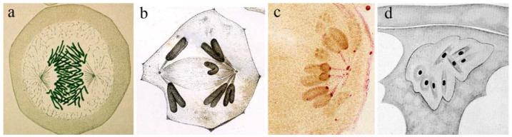
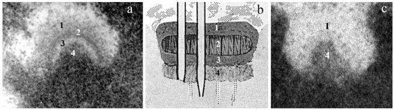
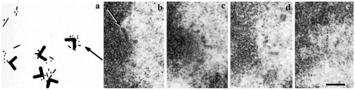
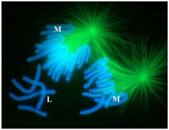
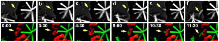
The attachment to and movement of a chromosome on the mitotic spindle are mediated by the formation of a bundle of microtubules (MTs) that tethers the kinetochore on the chromosome to a spindle pole. The origin of these "kinetochore fibers" (K fibers) has been investigated for over 125 years. As noted in 1944 by Schrader [Mitosis, Columbia University Press, New York, 110 pp.], there are three possible ways to form a K fiber: (a) it grows from the pole until it contacts the kinetochore, (b) it grows directly from the kinetochore, or (c) it forms as a result of an interaction between the pole and the chromosome. Since Schrader's time, it has been firmly established that K fibers in centrosome-containing animal somatic cells form as kinetochores capture MTs growing from the spindle pole (route a). It is now similarly clear that in cells lacking centrosomes, including higher plants and many animal oocytes, K fibers "self-assemble" from MTs generated by the chromosomes (route b). Can animal somatic cells form K fibers in the absence of centrosomes by the "self-assembly" pathway? In 2000, the answer to this question was shown to be a resounding "yes." With this result, the next question became whether the presence of a centrosome normally suppresses K fiber self-assembly or if this route works concurrently with centrosome-mediated K-fiber formation. This question, too, has recently been answered: observations on untreated live animal cells expressing green fluorescent protein-tagged tubulin clearly show that kinetochores can nucleate the formation of their associated MTs in a unique manner in the presence of functional centrosomes. The concurrent operation of these two "dueling" routes for forming K fibers in animal cells helps explain why the attachment of kinetochores and the maturation of K fibers occur as quickly as they do on all chromosomes within a cell.


Figures

Fig. 1
Fig. 1
Early evidence for kinetochores and K fibers. a Flemming's (1882) drawing of a newt cell in anaphase of mitosis. He did not observe in his preparations that each chromosome was connected to a spindle pole by a discrete fiber. b Druner's (1895) drawing showing K fibers during anaphase in an insect spermatocyte. c Metzner's (1894) drawing depicting kinetochores during anaphase. d Hughes-Schrader's (1924) illustration of prometaphase in A.wheeleri oocytes. Note that each chromosome appears to be organizing its own minispindle within an intact nuclear envelope
Fig. 2
Fig. 2
Mammalian kinetochore structure. a An unattached kinetochore from a newt cell viewed in a 0.25-μm section after conventional fixation, embedding, and staining. Note the extensive corona material (1) and the trilaminar structure of the kinetochore proper (2-4). b Jokelainen's (1967) early drawing of a kinetochore in a fetal rat cell. c An unattached kinetochore from a colcemid-treated PtK1 cell, viewed in a 0.25-μm section, after high-pressure freezing and freeze substitution. Numbers 1 and 4 note the corona and kinetochore proper, respectively. See text for details. (Micrograph in c is courtesy of B.F. McEwen and Y. Dong from the Wadsworth Center.)
Fig. 3
Fig. 3
Kinetochores nucleate MTs during a recovery from colcemid treatment. a Reconstruction from 33 serial 0.25-μm-thick sections of several kinetochores (chevron indicates kinetochore outer disk, chevron pointing away from the chromosome) in a CHO cell fixed 15 min into a recovery from a prolonged colcemid block. Note that numerous short MTs appear first near each kinetochore. b-e Four serial sections through the kinetochore depicted by the arrow in a. Note that numerous short MTs appear in the corona material (from Witt et al. 1980)
Fig. 4
Fig. 4
The kinetochores on newt chromosomes (blue) that are not incorporated into a forming spindle show no evidence of nucleating MTs (green). This indirect immunofluorescence micrograph of an early prometaphase newt cell contains several lost (L) and mono-oriented (M) chromosomes. Many of the mono-oriented chromosomes can clearly be seen to possess just a single K fiber. Note the absence of MTs in the vicinity of the lost chromosomes
Fig. 5
Fig. 5
Kinetochores in animal cells can nucleate MTs in the presence of a functional centrosome. a-f Frames from a fluorescence time-lapse recording of a Drosophila cell stably expressing GF/α-tubulin. The top frames show a deconvolved fluorescence image, while the bottom frames are an overlay of tubulin fluorescence (green) and chromosomes (red). The yellow arrow notes the nucleation and growth of MTs from an unattached kinetochore on a mono-oriented chromosome that is not facing a centrosome (from Maiato et al. 2004a)

References

    1. Bergen LG, Kuriyama R, Borisy GG. Polarity of microtubules nucleated by centrosomes and chromosomes of Chinese hamster ovary cells in vitro. J Cell Biol. 1980;84:151–159. - PMC - PubMed
    1. Biggins S, Walczak CE. Captivating capture: how microtubules attach to kinetochores. Curr Biol. 2003;13:R449–R460. - PubMed
    1. Brenner S, Pepper D, Berns MW, Tan E, Brinkley BR. Kinetochore structure, duplication, and distribution in mammalian cells: analysis by human autoantibodies from scleroderma patients. J Cell Biol. 1981;91:95–102. - PMC - PubMed
    1. Brinkley BR, Stubblefield E. The fine structure of the kinetochore of a mammalian cell in vitro. Chromosoma. 1966;19:28–43. - PubMed
    1. Carlson JG. Mitotic behavior of induced chromosomal fragments lacking spindle attachments in the neuroblasts of the grasshopper. Proc Natl Acad Sci U S A. 1938;24:500–507. - PMC - PubMed