. 2005 Aug 18;1(1):2.
doi: 10.1186/1746-4811-1-2.

Metabolic profiling of laser microdissected vascular bundles of Arabidopsis thaliana

Affiliations

Metabolic profiling of laser microdissected vascular bundles of Arabidopsis thaliana

Martina Schad et al. Plant Methods. .

Abstract

Background: Laser microdissection is a useful tool for collecting tissue-specific samples or even single cells from animal and plant tissue sections. This technique has been successfully employed to study cell type-specific expression at the RNA, and more recently also at the protein level. However, metabolites were not amenable to analysis after laser microdissection, due to the procedures routinely applied for sample preparation. Using standard tissue fixation and embedding protocols to prepare histological sections, metabolites are either efficiently extracted by dehydrating solvents, or washed out by embedding agents.

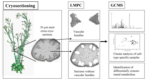
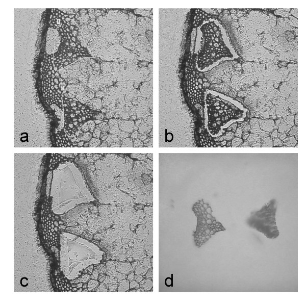
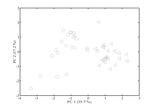
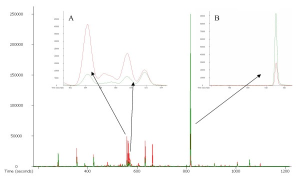
Results: In this study, we used cryosectioning as an alternative method that preserves sufficient cellular structure while minimizing metabolite loss by excluding any solute exchange steps. Using this pre-treatment procedure, Arabidopsis thaliana stem sections were prepared for laser microdissection of vascular bundles. Collected samples were subsequently analyzed by gas chromatography-time of flight mass spectrometry (GC-TOF MS) to obtain metabolite profiles. From 100 collected vascular bundles (approximately 5,000 cells), 68 metabolites could be identified. More than half of the identified metabolites could be shown to be enriched or depleted in vascular bundles as compared to the surrounding tissues.

Conclusion: This study uses the example of vascular bundles to demonstrate for the first time that it is possible to analyze a comprehensive set of metabolites from laser microdissected samples at a tissue-specific level, given that a suitable sample preparation procedure is used.


Figures

Figure 1
Figure 1
Experimental strategy.Outline of the strategy for tissue-specific metabolite profiling in A. thaliana. Stem cross cryosections were used for LMPC. Metabolites from microdissected vascular bundles and sections without vascular bundles, respectively, were extracted and analyzed by GCMS measurements.
Figure 2
Figure 2
Laser microdissection.The process of laser microdissection coupled to laser pressure catapulting (LMPC) for collecting vascular bundles from 30 μm thick cross sections obtained after freezing and cryosectioning A. thaliana stems. (a) Vascular bundles are selected on the computer screen. (b) The laser beam cuts along the markings. (c) The cut cells of interest are catapulted off the slide by a defocused laser pulse and (d) are collected into a cap of a microfuge tube.
Figure 3
Figure 3
Statistical evaluation of the metabolite data.Principal Components Analysis (PCA) of metabolite samples generated from A. thaliana stem tissue. The PCA score plot for principal component 1 (PC1) versus principal component 2 (PC2) is presented. The circles represent microdissected vascular bundle tissues (21 samples) whilst the triangles are samples from sections without vascular bundles (23 samples).
Figure 4
Figure 4
Example chromatogram.Entire chromatogram for m/z 217, an ion trace for sugars and sugar alcohols. The vascular bundle sample is shown in green and the sample without vascular bundle in red. (a) shows an example of depleted sugars in vascular bundles – fructose (retention time 564.19 s) and glucose (retention time 569.419 s) while (b) illustrates the zoomed-in sucrose peak (retention time 815.369 s) being enriched in vascular bundles.

References

    1. Tomos AD, Hinde P, Pritchard J, Fricke W. Microsampling and measurements of solutes in single cells. Plant cell biology: a practical approach Edited by Harris N, Oparka KJ Oxford: Oxford University Press. 1994. pp. 297–314.
    1. Andersson H, van den Berg A. Microtechnologies and nanotechnologies for single-cell analysis. Current Opinion in Biotechnology. 2004;15:44–49. doi: 10.1016/j.copbio.2004.01.004. - DOI - PubMed
    1. Brandt S, Kehr J, Walz C, Imlau A, Willmitzer L, Fisahn J. A rapid method for detection of plant gene transcripts from single epidermal, mesophyll and companion cells of intact leaves. Plant Journal. 1999;20:245–250. doi: 10.1046/j.1365-313x.1999.00583.x. - DOI - PubMed
    1. Arlt K, Brandt S, Kehr J. Amino acid analysis in five pooled single plant cell samples using capillary electrophoresis coupled to laser-induced fluorescence detection. Journal of Chromatography A. 2001;926:319–325. doi: 10.1016/S0021-9673(01)01052-4. - DOI - PubMed
    1. Tomos AD, Sharrock RA. Cell sampling and analysis (SiCSA): metabolites measured at single cell resolution. Journal of Experimental Botany. 2001;52:623–630. doi: 10.1093/jexbot/52.356.623. - DOI - PubMed