. 2005 Nov;96(11):774-83.
doi: 10.1111/j.1349-7006.2005.00111.x.

Characterization of human NSCLC cell line with innate etoposide-resistance mediated by cytoplasmic localization of topoisomerase II alpha

Affiliations

Characterization of human NSCLC cell line with innate etoposide-resistance mediated by cytoplasmic localization of topoisomerase II alpha

Bautista de Lucio et al. Cancer Sci. 2005 Nov.

Abstract

Topoisomerase (topo) II alpha is a target for many chemotherapeutic agents in clinical use. In tumor cells resistant to topo II poisons, there have been descriptions of quantitative and qualitative alterations involved in this enzyme. More recently, the cytoplasmic localization of topo II alpha has been described as a mechanism to confer drug resistance. Here, we report the characterization of a human non-small-cell lung cancer cell line, INER-37, which shows an innate resistance to etoposide. In this cell line, etoposide resistance was directly associated with the expression of topo II alpha resident mainly in the cytoplasmic region. At the molecular level, INER-37 cells carry on a heterozygous gene deletion, transcribing two different topo II alpha mRNAs: 4.8 kb and 2.0 kb. The bigger 4.8 kb mRNA (missing 1.3 kb of 3' mRNA and including the untranslated region) is translated into a truncated cytoplasmic protein of approximately 160 kDa. The protein truncation affects at least 96 amino acids in the COOH-terminal region where the more proximal bipartite nuclear localization signal is located. The INER-37 cell line is the first cancer cell line reported with an innate mutation affecting the 3'-end region of the topo II alpha gene that confers a cytoplasmic localization of the enzyme and therefore an increased resistance to etoposide.


Figures
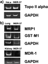
Figure 1
Figure 1
Reverse transcription polymerase chain reaction analysis of multidrug resistance (MDR)‐related genes on INER‐37 cells. Five micrograms of total RNA were used to synthesized cDNA and the fragments were amplified. (a) Topoisomerase II alpha expression in HeLa cells as a control and INER‐37 cells. (b) multidrug‐resistance related protein 1 (MRP‐1), glutathione S transferase (GST)‐M1 and MDR‐1 expression in INER‐37 cells. Lung and kidney tissues were used as a positive control of expression of GST‐M1, MRP1 and MDR‐1, respectively. The expression of the glyceraldehyde‐3 phosphate dehydrogenase (GAPDH) gene was used as a constitutive control for the integrity of the RNA molecules.
Figure 2
Figure 2
Catalytic Φ‐174 DNA unknotted‐mediated catalytic activity of topoisomerase II alpha activity. (a) Nuclear and (b) whole‐cell extracts from INER‐37 cells of HeLa cells were incubated with 0.5 µg of Φ‐174‐knotted DNA for 50 min. Samples were treated with proteinase K, sodium dodecylsulfate and electrophoresed on 1% agarose gel. (c) and (d) are densitometric graphics of three independent experiments showing the disappearing of the Φ‐174 DNA knotted form. Ctl, control Φ‐174‐knotted DNA.
Figure 3
Figure 3
Immunocytochemical detection of topo II alpha in INER‐37 and HeLa cell lines. Cells were seeded for 4 h in coverslides then probed with rabbit polyclonal anti‐topo II alpha antibody (kindly provided by Dr Leroy F. Liu), and immunostained by the peroxidase method using diaminobenzidine. Photomicrographs were taken with an optical microscope at ×400 magnification. (a) HeLa cells and (b–d) INER‐37 cells.
Figure 4
Figure 4
Representative molecular analysis of topoisomerase (topo) II alpha by reverse transcription polymerase chain reaction (RT‐PCR), rapid amplification of cDNA ends (3′‐RACE) and Northern blot. (a) Schematic representation of complete topo II alpha mRNA, recognizing the three major regions in the open reading frame as well as the untranslated region (3′‐UTR). The locations of the primers used for these studies are depicted as solid bars. (b) RT‐PCR analysis of the last 2 Kb of topo II alpha mRNA using the primers designed as fragment (F)I, FII and FIII (each sequence primer was taken from Campain et al. 1994) and the ATP‐primer was taken as a control. In all paragraphs, mRNA from HeLa cells (line 1) and INER‐37 cells (line 2) were analyzed. (c) The 3′‐RACE assay of the topo II alpha gene in INER‐37 cells. We analyzed the 3′‐end region of topo II alpha mRNA with 3′ RACE and nested RT‐PCR, using as sense a oligonucleotide matching in the 3857 nt and a primer matching with 17 bases of the RT‐primer used to translate the cDNA as antisense. (d) Northern blot analysis of topo II alpha in INER‐37 cells. Total RNA (10 µg) was separated by electrophoresis on a formaldehyde‐agarose denaturing gel and blotted onto nylon membrane. mRNAs of topo II alpha were detected with PCR cDNA probe (1246–1539 nt) obtained from HeLa cells.
Figure 5
Figure 5
Topoisomerase (topo) II alpha mRNA isoforms in cancer cell lines. Solid lines are the wild‐type sequences with respect to the full‐length topo II alpha from HeLa cells, and open bars are the new sequences identified in cancer cells lines. Nucleotide positions above bars indicate the mutation site in the altered transcript. Triangles are the sequences in the full‐length transcript but spliced out in the altered transcript.
Figure 6
Figure 6
Western blot analysis of whole extracts from INER‐37 and HeLa cells. Sixty micrograms of cell extract protein from INER‐37 and HeLa cells were subjected to sodium dodecylsulfate‐polyacrylamide gel electrophoresis, transferred to a nitrocellulose membrane, and probed with the following antibodies: two goat polyclonal anti‐topoisomerase (topo) II alpha antibodies raised against an internal epitope or recognizing an epitope mapping near the carboxyl terminus were worked at 1:100 dilution; a polyclonal antibody that recognizes the last 16 residues of the COOH‐terminal region of topo II alpha was used at 1:1000 dilution. To visualize the reactivity of the antibodies, a biotinylated swine anti‐goat‐mouse‐rabbit IgG, followed by enhanced chemiluminescence were used. Actin was used as the reference control.

References

    1. Wang JC. DNA topoisomerases. Annu Rev Biochem 1996; 65: 635–92. - PubMed
    1. Champoux JJ. DNA topoisomerases: Structure, function and mechanism. Annu Rev Biochem 2001; 70: 369–413. - PubMed
    1. Liu LF. DNA topoisomerase poisons as antitumor drugs. Annu Rev Biochem 1989; 58: 350–75. - PubMed
    1. Burden DA, Osheroff N. Mechanism of action of eukaryotic topoisomerase II and drugs targeted to the enzyme. Biochim Biophys Acta 1998; 1400: 139–54. - PubMed
    1. Bugg BY, Danks MK, Beck WT, Suttle DP. Expression of a mutant DNA topoisomerase II in CCRF‐CEM human leukemic cells selected for resistance to teniposide. Proc Natl Acad Sci USA 1991; 88: 7654–8. - PMC - PubMed

Publication types

MeSH terms