. 2006 Mar;34(3):293-304.
doi: 10.1165/rcmb.2005-0155OC. Epub 2005 Nov 4.

TNF-alpha sensitizes normal and fibrotic human lung fibroblasts to Fas-induced apoptosis

Affiliations

TNF-alpha sensitizes normal and fibrotic human lung fibroblasts to Fas-induced apoptosis

Stephen K Frankel et al. Am J Respir Cell Mol Biol. 2006 Mar.

Abstract

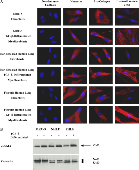
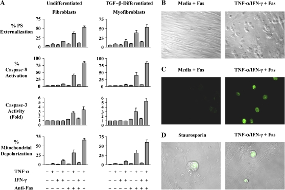
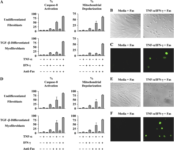
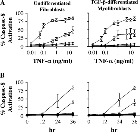
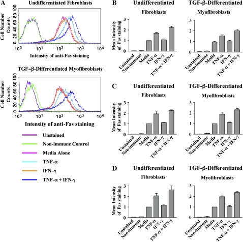
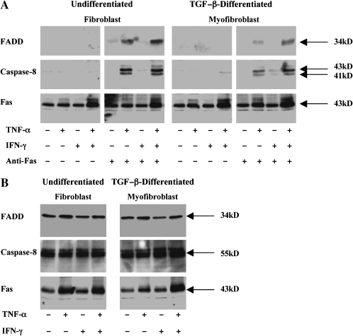
Pulmonary accumulation of fibroblasts and myofibroblasts in idiopathic pulmonary fibrosis/usual interstitial pneumonia (IFP/UIP) has been linked to (1) increased migration of a circulating pool of fibrocytes, (2) cell proliferation, and (3) resistance to apoptosis. The mechanism of physiologic apoptosis of lung fibroblasts is poorly understood. Using normal and fibrotic human lung fibroblasts and the human lung fibroblast cell line, MRC-5, we examined the regulation of Fas-induced apoptosis by the proinflammatory cytokines TNF-alpha and IFN-gamma. Herein, we show that the basal resistance of lung fibroblasts and myofibroblasts to Fas-induced apoptosis is overcome by sensitization with TNF-alpha. IFN-gamma did not sensitize cells to Fas-induced apoptosis, but exhibited synergistic activity with TNF-alpha. Sensitization by TNF-alpha was observed in MRC-5 cells and in fibroblasts and myofibroblasts from normal and fibrotic human lung, suggesting that this represents a conserved mechanism to engage Fas-induced apoptosis. The mechanism of sensitization was localized at the level of recruitment of the adapter protein, FADD, to the cytoplasmic domain of Fas. Collectively, these findings suggest that fibroblast apoptosis involves two steps, sensitization and induction, and that inadequate pulmonary inflammation in IPF/UIP may favor fibroblast accumulation by reducing sensitization to apoptosis.


Figures

<b>Figure 1.</b>
Figure 1.
Characterization of fibroblasts and myofibroblasts. (A) Immunocytochemistry for vimentin, pro-collagen I, and α-SMA expression in MRC-5, nondiseased human lung and fibrotic human lung fibroblast and TGF-β–differentiated myofibroblast cell populations. All fibroblast populations stained positive for vimentin and pro-collagen I with > 95% uniformity. All myofibroblast populations stained positive for vimentin, pro-collagen I, and α-SMA with > 95% uniformity and demonstrated organization of α-SMA into filaments. (B) Western blots showing relative expression of vimentin and α-SMA by MRC-5, nondiseased human lung and fibrotic human lung fibroblast and TGF-β–differentiated myofibroblast cell populations.
<b>Figure 2.</b>
Figure 2.
TNF-α–induced sensitization of MRC-5 fibroblasts and TGF-β–differentiated myofibroblasts to Fas-induced apoptosis. (A) MRC-5 fibroblasts and TGF-β–differentiated myofibroblasts were incubated in medium alone or with TNF-α (20 ng/ml), IFN-γ 50 (U/ml), or both cytokines for 36 h before challenge with agonistic anti-Fas antibody (250 ng/ml) for 6 h. The percentages of apoptotic cells were quantified by phosphatidylserine externalization, activation of caspases-8 and -3, and mitochondrial depolarization. The data shown represent the mean ± SD of at least three independent experiments. (B) Brightfield images of MRC-5 fibroblasts incubated with medium alone or TNF-α plus IFN-γ for 36 h before exposure to agonistic anti-Fas antibody for 6 h (magnification: ×10). (C) TUNEL staining of MRC-5 fibroblasts incubated with medium alone or TNF-α plus IFN-γ for 36 h before exposure to agonistic anti-Fas antibody for 6 h (magnification: ×63). (D) TUNEL staining superimposed on Nomarski images correlating morphologic changes of apoptosis with DNA cleavage. The MRC-5 fibroblasts pictured on the left were treated with 1 μM staurosporine for 12 h (positive control), whereas the cells on the right were treated with TNF-α plus IFN-γ for 36 h before exposure to agonistic anti-Fas antibody for 6 h.
<b>Figure 3.</b>
Figure 3.
(A) TNF-α–induced sensitization of normal human lung fibroblasts and TGF-β–differentiated myofibroblasts to Fas-induced apoptosis. Early passage normal human lung fibroblasts and TGF-β–differentiated (2 ng/ml, 72 h) myofibroblasts were incubated in medium alone or with TNF-α (20 ng/ml), IFN-γ (50 U/ml), or both cytokines for 48 h before exposure to agonistic anti-Fas antibody (250 ng/ml) for 12 h. The percentages of apoptotic cells were quantified by assays for caspase-8 activation and mitochondrial depolarization. The data shown represent the composite mean ± SD of fibroblasts derived from four donors each evaluated independently in triplicate. (B) Brightfield images of normal human lung fibroblasts incubated with medium alone or TNF-α plus IFN- γ for 48 h before exposure to agonistic anti-Fas anti-body for 12 h (magnification: ×10). (C) TUNEL staining of normal human lung fibroblasts incubated with medium alone or TNF-α plus IFN-γ for 48 h before exposure to agonistic anti-Fas antibody for 12 h (magnification: ×63). (D) TNF-α–induced sensitization of human fibrotic lung fibroblasts and TGF-β–differentiated myofibroblasts to Fas-induced apoptosis. Early passage human fibrotic lung fibroblasts and TGF-β–differentiated myofibroblasts were incubated in medium alone or with TNF-α (20 ng/ml), IFN-γ (50 U/ml), or both cytokines for 48 h before exposure to agonistic anti-Fas antibody (250 ng/ml) for 12 h. The percentages of apoptotic cells were quantified by assays for caspase-8 activation mitochondrial depolarization. The data shown represent the composite mean ± SD of fibroblasts derived from four donors each evaluated independently in triplicate. (E) Brightfield images of fibrotic human lung fibroblasts incubated with medium alone or TNF-α plus IFN- γ for 48 h before exposure to agonistic anti-Fas antibody for 12 h (magnification: ×10). (F) TUNEL staining of fibrotic human lung fibroblasts incubated with medium alone or TNF-α plus IFN-γ for 48 h before exposure to agonistic anti-Fas antibody for 12 h (magnification: ×63).
<b>Figure 4.</b>
Figure 4.
IFN-γ potentiates TNF-α sensitization to Fas-induced apoptosis in MRC-5 fibroblasts and TGF-β–differentiated myofibroblasts. (A) TNF-α dose response curve for MRC-5 fibroblasts and TGF-β–differentiated myofibroblasts. Cells were incubated with increasing doses of TNF-α (0.01–20 ng/ml) in the presence or absence of IFN-γ (50 U/ml) for 36 h followed by stimulation with agonistic anti-Fas antibody for 6 h. (B) Time course of sensitization of MRC-5 fibroblasts and TGF-β–differentiated myofibroblasts to Fas-induced apoptosis by TNF-α (20 ng/ml), IFN-γ (50 U/ml), or both cytokines. Squares, medium; triangles, TNF-α; inverted triangles, IFN-γ; diamonds, TNF-α + IFN- γ; filled symbols, no anti-Fas antibody; open symbols, with anti-Fas antibody. Apoptosis was quantified by caspase-8 activation.
<b>Figure 5.</b>
Figure 5.
Effect of TNF-α and IFN-γ on cell surface Fas expression. (A) MRC-5 fibroblasts and TGF-β–differentiated myofibroblasts were incubated in medium alone or with TNF-α (20 ng/ml), IFN-γ (50 U/ml), or both cytokines for 36 h before staining with APC-labeled anti-Fas antibody and analysis by flow cytometry. Representative flow cytometry histograms for Fas cell surface expression, repeated independently three times, are depicted. (B) Mean intensity of staining ± SEM of Fas expression levels. (C) Human lung fibroblasts and TGF-β–differentiated myofibroblasts were incubated in medium alone or with TNF-α (20 ng/ml), IFN-γ (50 U/ml), or both cytokines for 48 h before staining with APC-labeled anti-Fas antibody and analysis by flow cytometry. Mean intensity of staining ± SEM of Fas expression levels of pooled data with fibroblasts obtained from four normal lung donors, with cells from each individual donor repeated independently three times. (D) Mean intensity of staining ± SEM of Fas expression levels of pooled data with fibroblasts obtained from four fibrotic lung donors, with cells from each individual donor repeated independently three times.
<b>Figure 6.</b>
Figure 6.
Effect of TNF-α and IFN-γ on the assembly of the Fas-induced death inducing signaling complex (DISC). (A) MRC-5 fibroblasts and TGF-β–differentiated myofibroblasts were incubated in medium alone or with TNF-α (20 ng/ml), IFN-γ (50 U/ml), or both cytokines for 36 h before challenge with agonistic anti-Fas antibody (250 ng/ml) for 4 h. Cells were lysed and equal amounts of total cell protein were immunoprecipitated with anti-Fas antibody, washed and separated by SDS-PAGE. Co-immunoprecipitating FADD (34 kD) and active caspase-8 (41 and 43 kD) were detected by Western blotting with specific antibodies. Immunoprecipation of Fas (43 kD) was confirmed by stripping the blots and re-probing with anti-Fas antibody. (B) Western blot analysis of total FADD, caspase-8 and Fas levels in undifferentiated and TGF-β–differentiated MRC-5 cells. Cells were stimulated as in A., lysed, and equal amounts of total cell lysate proteins were analyzed by SDS-PAGE and Western blotting with the appropriate antibody. Data shown are representative results of three independent experiments.
<b>Figure 7.</b>
Figure 7.
(A) Nondiseased human lung fibroblasts and TGF-β–differentiated myofibroblasts were incubated in medium alone or with TNF-α (20 ng/ml), IFN-γ (50 U/ml), or both cytokines for 48 h before challenge with agonistic anti-Fas antibody (250 ng/ml) for 6 h Cells were lysed and analyzed as described in the legend to Figure 6. Data shown are representative results of three independent experiments for each of four patients. (B) Western blot analysis of total FADD, caspase-8, and Fas levels in undifferentiated and TGF-β–differentiated nondiseased human lung cells. Data shown are representative result of three independent experiments for each of four patients.
<b>Figure 8.</b>
Figure 8.
(A) Fibrotic human lung fibroblasts and TGF-β–differentiated myofibroblasts were incubated in medium alone or with TNF-α (20 ng/ml), IFN-γ (50 U/ml), or both cytokines for 48 h before challenge with agonistic anti-Fas antibody (250 ng/ml) for 6 h. Cells were lysed and analyzed as described in the legend to Figure 6. Data shown are representative results of three independent experiments for each of four patients. (B) Western blot analysis of total FADD, caspase-8, and Fas levels in undifferentiated and TGF-β–differentiated fibrotic human lung cells. Data shown are representative results of three independent experiments for each of four patients.

References

    1. American Thoracic Society. Idiopathic pulmonary fibrosis: diagnosis and treatment. International consensus statement. American Thoracic Society (ATS), and the European Respiratory Society (ERS). Am J Respir Crit Care Med 2000;161:646–664. - PubMed
    1. Myers JL, Katzenstein AL. Epithelial necrosis and alveolar collapse in the pathogenesis of usual interstitial pneumonia. Chest 1988;94:1309–1311. - PubMed
    1. Kuhn C III, Boldt J, King TE Jr, Crouch E, Vartio T, McDonald JA. An immunohistochemical study of architectural remodeling and connective tissue synthesis in pulmonary fibrosis. Am Rev Respir Dis 1989;140:1693–1703. - PubMed
    1. Kuhn C, McDonald JA. The roles of the myofibroblast in idiopathic pulmonary fibrosis. Ultrastructural and immunohistochemical features of sites of active extracellular matrix synthesis. Am J Pathol 1991;138:1257–1265. - PMC - PubMed
    1. Hashimoto N, Jin H, Liu T, Chensue SW, Phan SH. Bone marrow-derived progenitor cells in pulmonary fibrosis. J Clin Invest 2004;113:243–252. - PMC - PubMed

Publication types

MeSH terms