Characterization of a serodiagnostic complement fixation antigen of Coccidioides posadasii expressed in the nonpathogenic Fungus Uncinocarpus reesii
- PMID: 16272471
- PMCID: PMC1287831
- DOI: 10.1128/JCM.43.11.5462-5469.2005
Characterization of a serodiagnostic complement fixation antigen of Coccidioides posadasii expressed in the nonpathogenic Fungus Uncinocarpus reesii
Abstract
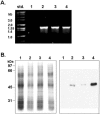
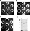
Coccidioides spp. (immitis and posadasii) are the causative agents of human coccidioidomycosis. In this study, we developed a novel system to overexpress coccidioidal proteins in a nonpathogenic fungus, Uncinocarpus reesii, which is closely related to Coccidioides. A promoter derived from the heat shock protein gene (HSP60) of Coccidioides posadasii was used to control the transcription of the inserted gene in the constructed coccidioidal protein expression vector (pCE). The chitinase gene (CTS1) of C. posadasii, which encodes the complement fixation antigen, was expressed using this system. The recombinant Cts1 protein (rCts1(Ur)) was induced in pCE-CTS1-transformed U. reesii by elevating the cultivation temperature. The isolated rCts1(Ur) showed chitinolytic activity that was identical to that of the native protein and had serodiagnostic efficacy comparable to those of the commercially available antigens in immunodiffusion-complement fixation tests. Using the purified rCts1(Ur), 74 out of the 77 coccidioidomycosis patients examined (96.1%) were positively identified by enzyme-linked immunosorbent assay. The rCts1(Ur) protein showed higher chitinolytic activity and slightly greater seroreactivity than the bacterially expressed recombinant Cts1. These data suggest that this novel expression system is a useful tool to produce coccidioidal antigens for use as diagnostic antigens.
Figures


Similar articles
-
Characterization of an Uncinocarpus reesii-expressed recombinant tube precipitin antigen of Coccidioides posadasii for serodiagnosis.PLoS One. 2019 Aug 14;14(8):e0221228. doi: 10.1371/journal.pone.0221228. eCollection 2019. PLoS One. 2019. PMID: 31412087 Free PMC article. Clinical Trial.
-
Disruption of the gene which encodes a serodiagnostic antigen and chitinase of the human fungal pathogen Coccidioides immitis.Infect Immun. 2000 Oct;68(10):5830-8. doi: 10.1128/IAI.68.10.5830-5838.2000. Infect Immun. 2000. PMID: 10992492 Free PMC article.
-
The coccidioidal complement fixation and immunodiffusion-complement fixation antigen is a chitinase.Infect Immun. 1992 Jul;60(7):2588-92. doi: 10.1128/iai.60.7.2588-2592.1992. Infect Immun. 1992. PMID: 1612728 Free PMC article.
-
Laboratory aspects in the diagnosis of coccidioidomycosis.Ann N Y Acad Sci. 2007 Sep;1111:301-14. doi: 10.1196/annals.1406.049. Epub 2007 Mar 15. Ann N Y Acad Sci. 2007. PMID: 17363434 Review.
-
Pulmonary coccidioidomycosis.Semin Respir Crit Care Med. 2011 Dec;32(6):754-63. doi: 10.1055/s-0031-1295723. Epub 2011 Dec 13. Semin Respir Crit Care Med. 2011. PMID: 22167403 Review.
Cited by
-
A Recombinant Multivalent Vaccine (rCpa1) Induces Protection for C57BL/6 and HLA Transgenic Mice against Pulmonary Infection with Both Species of Coccidioides.Vaccines (Basel). 2024 Jan 9;12(1):67. doi: 10.3390/vaccines12010067. Vaccines (Basel). 2024. PMID: 38250880 Free PMC article.
-
Multivalent recombinant protein vaccine against coccidioidomycosis.Infect Immun. 2006 Oct;74(10):5802-13. doi: 10.1128/IAI.00961-06. Infect Immun. 2006. PMID: 16988258 Free PMC article.
-
The Quest for a Vaccine Against Coccidioidomycosis: A Neglected Disease of the Americas.J Fungi (Basel). 2016 Dec 16;2(4):34. doi: 10.3390/jof2040034. J Fungi (Basel). 2016. PMID: 29376949 Free PMC article. Review.
-
Characterization of an Uncinocarpus reesii-expressed recombinant tube precipitin antigen of Coccidioides posadasii for serodiagnosis.PLoS One. 2019 Aug 14;14(8):e0221228. doi: 10.1371/journal.pone.0221228. eCollection 2019. PLoS One. 2019. PMID: 31412087 Free PMC article. Clinical Trial.
-
Two lateral flow assays for detection of anti-coccidioidal antibodies show similar performance to immunodiffusion in dogs with coccidioidomycosis.Am J Vet Res. 2024 Mar 25;85(6):ajvr.23.12.0272. doi: 10.2460/ajvr.23.12.0272. Print 2024 Jun 1. Am J Vet Res. 2024. PMID: 38531155 Free PMC article.
References
-
- Bowman, B. H., T. J. White, and J. W. Taylor. 1996. Human pathogenic fungi and their close nonpathogenic relatives. Mol. Phylogenet. Evol. 6:89-96. - PubMed
-
- Coligan, J., B. Dunn, H. Ploegh, D. Speicher, and P. Wingfield. 1995. Current protocols in protein science. John Wiley & Sons Inc., New York, N.Y.
Publication types
MeSH terms
Substances
Grants and funding
LinkOut - more resources
Full Text Sources
Other Literature Sources
Medical
Research Materials
Miscellaneous

