. 2005 Dec;7(12):1240-7.
doi: 10.1038/ncb1335. Epub 2005 Nov 6.

RNA-dependent integrin alpha3 protein localization regulated by the Muscleblind-like protein MLP1

Affiliations

RNA-dependent integrin alpha3 protein localization regulated by the Muscleblind-like protein MLP1

Yair Adereth et al. Nat Cell Biol. 2005 Dec.

Erratum in

  • Nat Cell Biol. 2006 Jan;8(1):100

Abstract

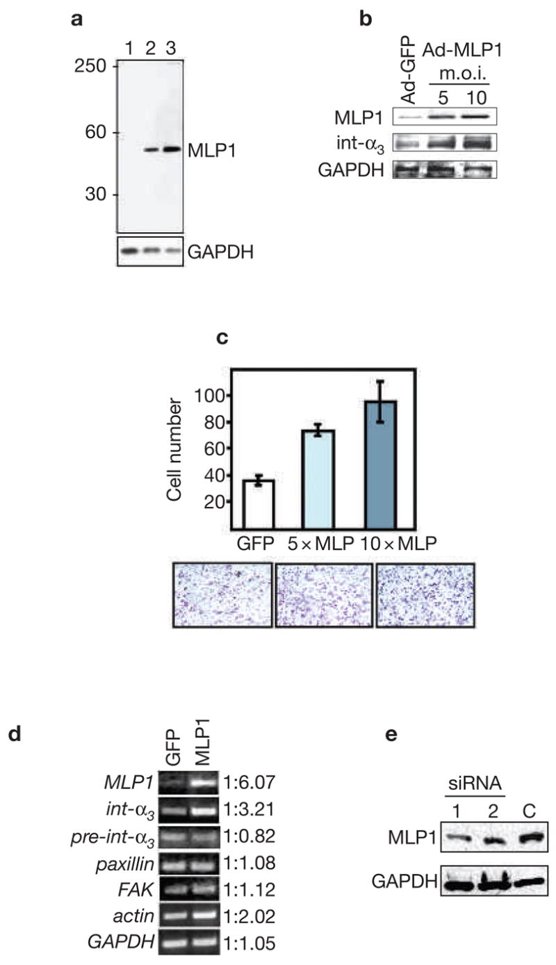
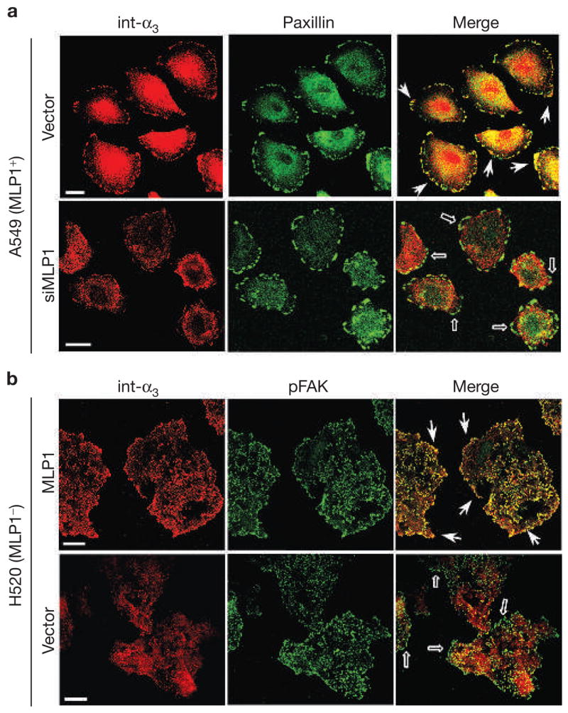
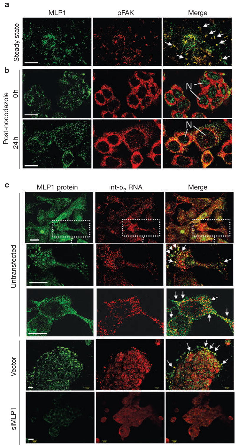
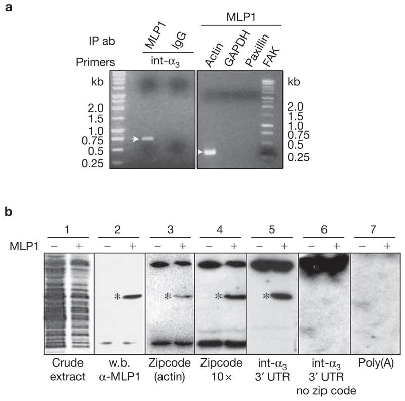
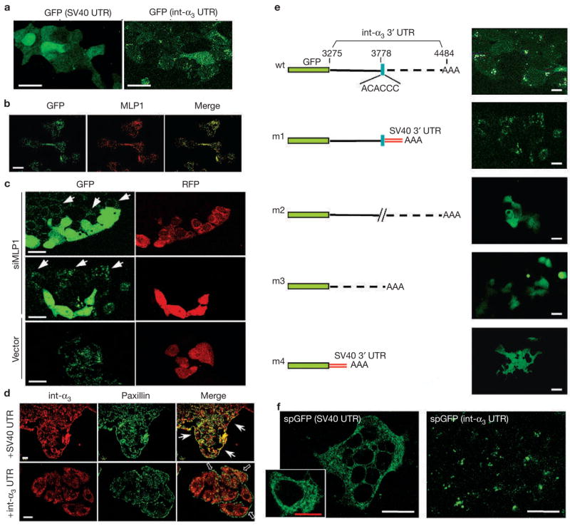
We show that localized expression of the integrin alpha3 protein is regulated at the level of RNA localization by the human homologue of Drosophila Muscleblind, MLP1/MBLL/MBNL2, a unique Cys3His zinc-finger protein. This is supported by the following observations: MLP1 knockdown abolishes localization of integrin alpha3 to the adhesion complexes; MLP1 is localized in adhesion plaques that contain phospho-focal adhesion kinase; this localization is microtubule-dependent; integrin alpha3 transcripts colocalize with MLP1 in distinct cytoplasmic loci; integrin alpha3 transcripts are physically associated with MLP1 in cells and MLP1 binds to a specific ACACCC motif in the integrin alpha3 3' untranslated region (UTR) in vitro; and a green fluorescent protein (GFP) open reading frame-integrin alpha3 3' UTR chimeric gene directs GFP protein localization to distinct cytoplasmic loci near the cell periphery, which is dependent on MLP1 and is mediated by the ACACCC motif but is independent of the integrin alpha3 signal peptide.


Figures

Figure 1
Figure 1
MLP1 regulates integrin α3 expression at the post-transcriptional level. (a) Protein extracts (10 μg/lane) from three cell lines were probed with rabbit anti-MLP1 antibody. The blot was stripped and re-probed for glyceraldehyde-3-phosphate dehydrogenase (GAPDH) as the loading control. Lane 1: H520 cells; lane 2: A549 cells; lane 3: HT29 cells. A single band of expected size (relative molecular mass of approximately 40,000) was detected. MLP1 was not detectable in H520 cells with the amount of total protein extracts used. (b) H520 cells were infected with empty adenoviral vector (Ad-GFP; m.o.i. 10) or vector containing the MLP1 coding sequence (Ad-MLP1) at m.o.i. of 5 or 10. Total protein extracts (50 μg; higher amount needed for detecting integrins) were probed with the antibodies indicated on the left. GAPDH served as the loading control. Increased expression of MLP1 correlates with increased expression of integrin α3. (c) H520 cells were transfected with adenoviral vector expressing green fluorescent protein (GFP) alone (at 10× m.o.i.), or GFP plus MLP1 (at 5× and 10× m.o.i.), and plated onto the upper chamber of the transwell. Cells that migrated to the underside of the upper chamber were stained with crystal violet and the numbers compared. Lower panels show representative views of the migrated cells. (d) Reverse transcription polymerase chain reaction (RT-PCR) analysis using primers specific for MLP1 mRNA, integrin α3 exon (int-α3), integrin α3 intron–exon junction (pre-int-α3), or mRNAs of paxillin, focal adhesion kinase (FAK), actin and GAPDH. Total RNAs were isolated from H520 cells infected with adenoviral vector (GFP) or vector containing the MLP1 coding sequence at 5× m.o.i. The linear range of PCR amplification was empirically determined for each primer set (see Methods). At the mid-point of the linear curve, amplified samples were run on ethidium-bromide-stained agarose gel and quantified. Integrin α3 RNA level increases in step with increased expression of MLP1, whereas the pre-spliced integrin α3 RNA level remains unchanged. There is a moderate increase of actin in the presence of MLP1 and no change for paxillin, FAK or GAPDH. Note that without reverse transcriptase, the PCR reactions did not yield any amplified products (data not shown). (e) A549 cells were transfected with empty vector (C) or vector containing one of the two MLP1-specific small interfering RNA (siRNA)-encoding oligonucleotide duplexes.
Figure 2
Figure 2
MLP1 is necessary for integrin α3 protein localization to adhesion plaques. (a) A549 cells transfected with empty vector or vector containing MLP1-specific oligonucleotide duplex #1 (siMLP1), as in Fig. 1e, were double-stained for paxillin (green) and integrin α3 (int-α3; red). In vector-transfected cells, integrin α3 colocalized prominently with paxillin in adhesion plaques at the migration front (examples indicated by arrows). In cells transfected with vector expressing MLP1-specific small interfering RNA (siRNA), integrin α3 no longer colocalized with paxillin in the cell periphery (empty arrows). (b) H520 cells were infected with empty adenovector or vector containing the MLP1 coding sequence at the m.o.i. of 10 (as in Fig. 1b), and double-stained for phospho-focal adhesion kinase (pFAK; green) and integrin α3 (red). Each stained area is a cluster of 5–10 cells but the peripheral pFAK appears mostly in cells at the leading edge. Without MLP1, integrin α3 is not colocalized with pFAK at the periphery (empty arrows). Expression of MLP1 results in re-localization of integrin α3 to pFAK-containing plaques at the periphery (arrows). Scale bars, 20 μm.
Figure 3
Figure 3
MLP1 regulates localization of integrin α3 RNA. (a) A549 cells were double-stained for MLP1 (green) and phospho-focal adhesion kinase (pFAK; red). Arrows mark examples of prominent adhesion plaques, where MLP1 and pFAK colocalize. (b) HT29 cells were treated with nocodazole and then returned to normal media. Cells were fixed and double-stained for MLP1 (green) and pFAK (red) at 0 and 24 h after recovery. Before recovery from nocodazole treatment (0 h), MLP1 was mainly localized in the nuclei (N). Twenty-four hours after recovery, MLP1 distribution returned to a punctated pattern with substantial colocalization with pFAK. (c) HT29 cells were double-stained for MLP1 protein and integrin α3 (int-α3) RNA. Untransfected cells and cells transfected with empty vector or vector expressing MLP1 small interfering RNA (siRNA) are indicated. In untransfected samples, a cell cluster and two enlarged single-cell views are shown to better visualize the large complexes that have been co-stained for integrin α3 RNA and MLP1 protein. In vector and siMLP1-transfected samples, clusters of cells are shown and the integrin α3 RNA–MLP1-containing complexes are mostly at the leading edges (arrows). Sense integrin α3 RNA probe showed no signals (data not shown). Scale bars, 20 μm.
Figure 4
Figure 4
MLP1 interacts with integrin α3 3′ untranslated region (UTR) in vivo and in vitro. (a) H520 cells were transfected with 10× m.o.i. of adenovirus-containing MLP1 coding sequence and total RNAs were subjected to RNA pulldown and reverse transcription polymerase chain reaction (RT-PCR). Integrin α3 (int-α3) RNA was detected in the fraction that was pulled down by MLP1 antibody, yielding an expected fragment size (see Methods) on an ethidium-bromide-stained agarose gel. Pulldown by purified rabbit immunoglobulin G (IgG) served as a negative control. In addition, actin mRNA was present in the MLP1-containing complex, which was detected by RT-PCR yielding the expected 337-bp fragment, whereas paxillin, focal adhesion protein (FAK) and glyceraldehyde-3-phosphate dehydrogenase (GAPDH) were not detectable. IP ab, immunoprecipitated antibodies (b) Extracts from BL21<DE3> bacteria transformed with (+) or without (−) plasmids containing MLP1 were run on SDS–PAGE and stained with Coomassie brilliant blue (blot 1) or western-blotted (w.b.) with anti-MLP1 antibody (blot 2). The MLP1 protein band is marked (*). Duplicate lanes were transferred to nitrocellulose membrane, renatured and hybridized with various 32P-labelled RNAs indicated at the bottom (blots 3–7). MLP1 (*) binds only to RNA molecules containing the zipcode (blots 3, 4 and 5). The bands at the top and bottom of the gels, present in both MLP1+ and MLP1 samples, were probably bacterial RNA-binding proteins.
Figure 5
Figure 5
Localized GFP expression mediated by the zipcode in integrin α3 3′ UTR. (a) HT29 cells were transfected with vectors containing enhanced green fluorescent protein (eGFP) open reading frame upstream of the SV40 3′ untranslated region (UTR) or the integrin α3 (int-α3) 3′UTR, and observed for GFP expression. With SV40 3′ UTR, GFP showed a diffuse expression pattern throughout the cell body. With integrin α3 3′ UTR, GFP was localized in a punctated pattern. (b) The same HT29 cells that were transfected with GFP–integrin-α3 chimaeras were fixed and stained for GFP and MLP1. The two proteins colocalized in subcellular punctates. (c) HT29 cells expressing GFP–integrin-α3 chimaeras were transfected with vector containing red fluorescent protein (RFP) alone (vector) or RFP and MLP1 small interfering RNA (siRNA) #1 (siMLP1; see Fig. 1). Live cells were examined. In the presence of siMLP1 (co-expressing RFP) the GFP became diffuse, whereas in neighbouring untransfected cells (arrows) the GFP remained in a punctated pattern. Transfection by the vector alone did not have any effects on the punctated GFP expression. (d) Transfected HT29 cells (+SV40 UTR or +int-α3 UTR) were stained for the endogenous integrin α3 (red) and paxillin (green). Ectopic overexpression of integrin α3 3′ UTR resulted in dispersion of endogenous integrin α3 protein from the paxillin-containing plaques (empty arrows), whereas overexpression of SV40 3′ UTR had little effect. Arrows point to plaques containing integrin α3 and paxillin at the leading edges. (e) Various deletion constructs of the integrin α3 3′ UTR were tested for the essential cis-acting localization element. AAA depicts the polyadenylation signal. SV40 3′ UTR that was contained in the cloning vector provided the default polyadenylation signal for the m1 and m4 constructs. The zipcode (blue box) seemed to be the most critical cis element. wt, wild type. (f) Chimeric integrin α3 signal peptide (sp)-GFP–SV40 or spGFP–integrin-α3 3′ UTR were transfected into HT29 cells and the GFP patterns were observed. With SV40 3′ UTR, the expected membrane-targeted GFP (spGFP) appears associated with an intracellular network, presumably the ER. The inset shows an enlarged view of a single cell displaying the network-bound GFP. With the integrin α3 3′ UTR, the GFP remains in the same punctated pattern as without the signal peptide. Scale bars, 20 μm, except the red bar in the inset, which is 10 μm.

References

    1. Begemann G, Paricio N, Artero R, Kiss I, Perez-Alonso M, Mlodzik M. Muscleblind, a gene required for photoreceptor differentiation in Drosophila, encodes novel nuclear Cys3His-type zinc-finger-containing proteins. Development. 1997;124:4321–4331. - PubMed
    1. Artero R, et al. The muscleblind gene participates in the organization of Z-bands and epidermal attachments of Drosophila muscles and is regulated by Dmef2. Dev Biol. 1998;195:131–143. - PubMed
    1. Miller JW, et al. Recruitment of human muscleblind proteins to (CUG)(n) expansions associated with myotonic dystrophy. EMBO J. 2000;19:4439–4448. - PMC - PubMed
    1. Fardaei M, Larkin K, Brook JD, Hamshere MG. In vivo co-localisation of MBNL protein with DMPK expanded-repeat transcripts. Nucleic Acids Res. 2001;29:2766–2771. - PMC - PubMed
    1. Fardaei M, et al. Three proteins, MBNL, MBLL and MBXL, co-localize in vivo with nuclear foci of expanded-repeat transcripts in DM1 and DM2 cells. Hum Mol Genet. 2002;11:805–814. - PubMed

Publication types