. 2005 Dec;12(12):1086-93.
doi: 10.1038/nsmb1015. Epub 2005 Nov 6.

Structural basis for transcription inhibition by tagetitoxin

Affiliations

Structural basis for transcription inhibition by tagetitoxin

Dmitry G Vassylyev et al. Nat Struct Mol Biol. 2005 Dec.

Abstract

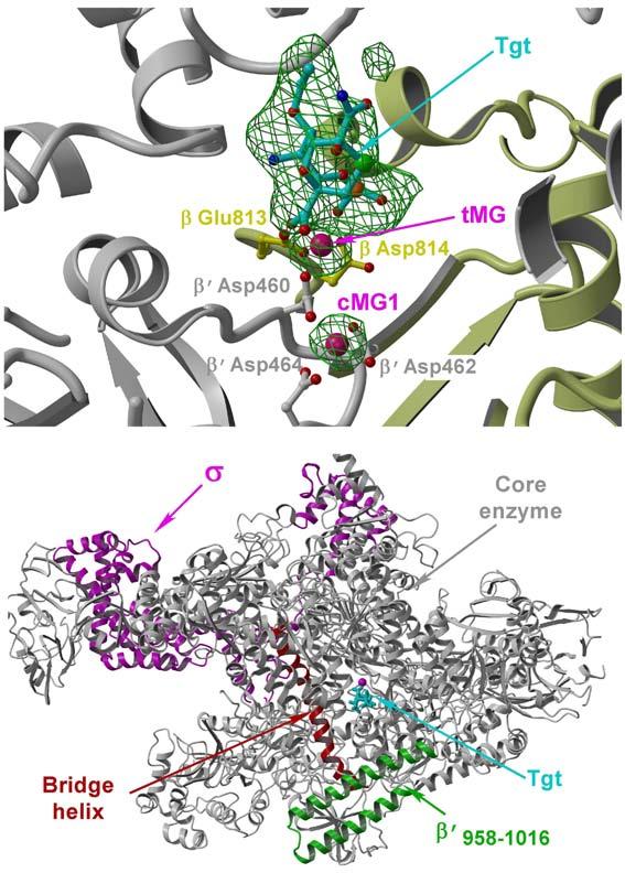
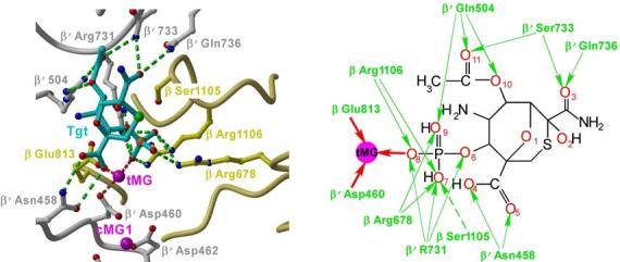
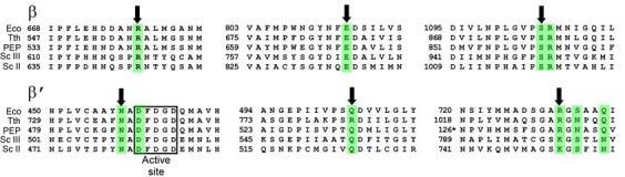
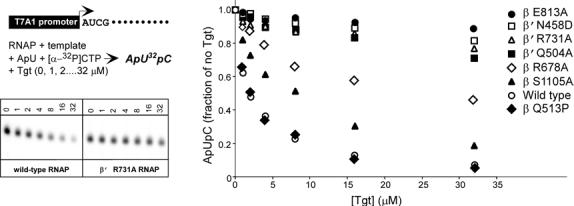
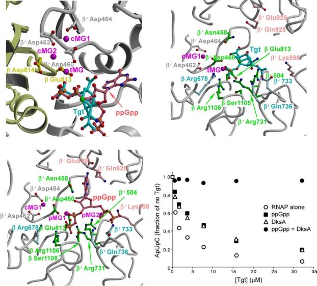
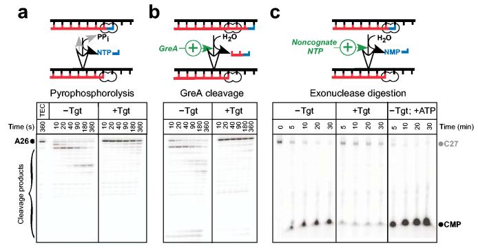
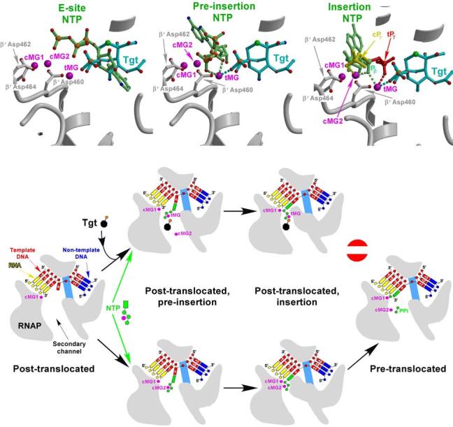
Tagetitoxin (Tgt) inhibits transcription by an unknown mechanism. A structure at a resolution of 2.4 A of the Thermus thermophilus RNA polymerase (RNAP)-Tgt complex revealed that the Tgt-binding site within the RNAP secondary channel overlaps that of the stringent control effector ppGpp, which partially protects RNAP from Tgt inhibition. Tgt binding is mediated exclusively through polar interactions with the beta and beta' residues whose substitutions confer resistance to Tgt in vitro. Importantly, a Tgt phosphate, together with two active site acidic residues, coordinates the third Mg(2+) ion, which is distinct from the two catalytic metal ions. We show that Tgt inhibits all RNAP catalytic reactions and propose a mechanism in which the Tgt-bound Mg(2+) ion has a key role in stabilization of an inactive transcription intermediate. Remodeling of the active site by metal ions could be a common theme in the regulation of catalysis by nucleic acid enzymes.


Figures

Figure 1
Figure 1
RNAP/Tgt structure. (a) Experimental 3.5Å resolution and |FTgt - Fnati| (3.0σ level both) omit ED map (green) superimposed on the RNAP1/Tgt structure. The residues (balls-and-sticks) and protein backbone (ribbon diagram) of the β and β′ subunits are shown in yellow and gray, respectively. In this and following figures, cMG1, cMG2, and tMG designate high- and low-affinity catalytic and Tgt-bound Mg2+ ions, respectively. (b) Overall view of the RNAP/Tgt complex structure showing that Tgt binds within the secondary channel in close vicinity to the active site (marked by cMG1, magenta sphere).
Figure 2
Figure 2
The Tgt-binding site. (a,b) A model (a) and schematic drawing (b) of the Tgt-binding site on RNAP. Residues that are not identical between E. coli and T. thermophilus are marked by their numbers only. Polar interactions are in red (tMG coordination bonds) or green (hydrogen bonds) dashed lines (a) and arrows (b). Weak contacts of β Ser1105 with Tgt are represented by a dashed arrow (b). (c) Sequence alignment of the β and β′ subunits from bacterial (E. coli, Eco; T. thermophilus, Tth), chloroplast (Arabidopsis thaliana, Ath), and yeast Saccharomyces cerevisiae (pol II, Sc II; and pol III, Sc III) enzymes in regions flanking the Tgt contact sites using DNAStar MegAlign Module. The Tgt structural determinants are highlighted by green boxes; residues substituted for the in vitro analysis are indicated by black arrows. (d) Inhibition of abortive transcription on the T7A1 promoter by Tgt. Formation of the radiolabeled ApUpC RNAs was followed as a function of Tgt concentration (from 0 to 32 μM) with wild-type or altered ecRNAP. The key is shown in the figure.
Figure 2
Figure 2
The Tgt-binding site. (a,b) A model (a) and schematic drawing (b) of the Tgt-binding site on RNAP. Residues that are not identical between E. coli and T. thermophilus are marked by their numbers only. Polar interactions are in red (tMG coordination bonds) or green (hydrogen bonds) dashed lines (a) and arrows (b). Weak contacts of β Ser1105 with Tgt are represented by a dashed arrow (b). (c) Sequence alignment of the β and β′ subunits from bacterial (E. coli, Eco; T. thermophilus, Tth), chloroplast (Arabidopsis thaliana, Ath), and yeast Saccharomyces cerevisiae (pol II, Sc II; and pol III, Sc III) enzymes in regions flanking the Tgt contact sites using DNAStar MegAlign Module. The Tgt structural determinants are highlighted by green boxes; residues substituted for the in vitro analysis are indicated by black arrows. (d) Inhibition of abortive transcription on the T7A1 promoter by Tgt. Formation of the radiolabeled ApUpC RNAs was followed as a function of Tgt concentration (from 0 to 32 μM) with wild-type or altered ecRNAP. The key is shown in the figure.
Figure 3
Figure 3
Tgt and ppGpp bind to overlapping sites on RNAP. (a) Superposition of the ttRNAP/Tgt and ttRNAP/ppGpp complexes. The color scheme is the same as in Figures 1a and 2a. (b,c) Structural determinants likely crucial for the Tgt and ppGpp binding. The RNAP/Tgt (b) and RNAP/ppGpp (c) complexes are shown in the same orientation for better comparison. RNAP residues (balls-and-sticks) that interact with both Tgt and ppGpp are shown in green, whereas those specific for Tgt and ppGpp are shown in light cyan and light pink, respectively. (d) ppGpp and DksA compete with Tgt for the inhibition of abortive transcription by the wild-type ecRNAP from the T7A1 promoter. The assay was performed as in Figure 2d. ppGpp was added to 0.5 mM, DksA – to 500 nM.
Figure 4
Figure 4
Tgt inhibits all catalytic reactions. (a,b) Pyrophosphorolysis and GreA-enhanced transcript cleavage assays. Halted radiolabeled A26 TECs were purified from NTPs using G50 spin columns and pre-incubated with Tgt at 37 °C for 2 min. GreA (300 nM) or PPi (400 μM) were added at time 0, followed by incubation at 37 °C. (c) Exonuclease cleavage assay. 3′ radiolabeled C27 complexes were incubated with water (left panel), 2 μM Tgt (center), or 1 mM ATP (right panel) at 37 °C. Aliquots were removed at the indicated times and quenched.
Figure 5
Figure 5
Mechanism of Tgt action. (a,b,c) Models of the substrates (light green) corresponding to the three postulated consecutive steps during NTP loading to the RNAP active site are superimposed on the RNAP/Tgt structure; the E-site (a), the pre-insertion site (b), and the insertion site (c); the PDB accession codes used in panels a, b and c were 1R9T, 1Y77, and 1R9S, respectively. The putative coordination bonds with tMG and/or cMG2 of Tgt (cyan), the NTP in the pre-insertion site and the β-phosphate (Pβ) of the NTP in the insertion site (light green) (b,c), as well as of the NTP γ-phosphates in the insertion site (c) in the “catalytic” cPγ (yellow) and “inactive”, tMG-bound tPγ (red) configurations are shown by dashed lines. (d) Stabilization of an inactive transcription intermediate by Tgt.

Similar articles

Cited by

References

    1. Rozovskaia T, Chenchik A, Bibilashvili R. Reaction of pyrophosphorolysis catalyzed by Escherichia coli RNA polymerase. Mol. Biol. (Mosk) 1981;15:636–652. - PubMed
    1. Sosunov V, et al. Unified two-metal mechanism of RNA synthesis and degradation by RNA polymerase. EMBO J. 2003;22:2234–44. - PMC - PubMed
    1. Komissarova N, Kashlev M. Transcriptional arrest: Escherichia coli RNA polymerase translocates backward, leaving the 3' end of the RNA intact and extruded. Proc Natl Acad Sci U S A. 1997;94:1755–60. - PMC - PubMed
    1. Orlova M, et al. Intrinsic transcript cleavage activity of RNA polymerase. Proc. Natl. Acad. Sci. USA. 1995;92:4596–4600. - PMC - PubMed
    1. Steitz TA. A mechanism for all polymerases. Nature. 1998;391:231–2. - PubMed

Publication types

MeSH terms

Associated data

LinkOut - more resources