Clinical Trial
. 2006 Sep;55(9):1122-31.
doi: 10.1007/s00262-005-0089-3. Epub 2005 Nov 5.

NK cell activation by dendritic cell vaccine: a mechanism of action for clinical activity

Affiliations
Clinical Trial

NK cell activation by dendritic cell vaccine: a mechanism of action for clinical activity

Takuya Osada et al. Cancer Immunol Immunother. 2006 Sep.

Abstract

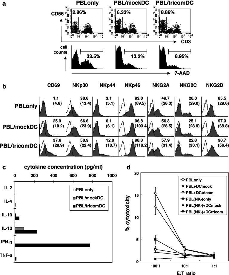
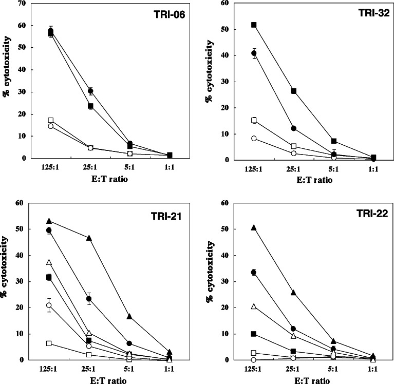
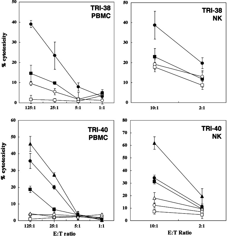
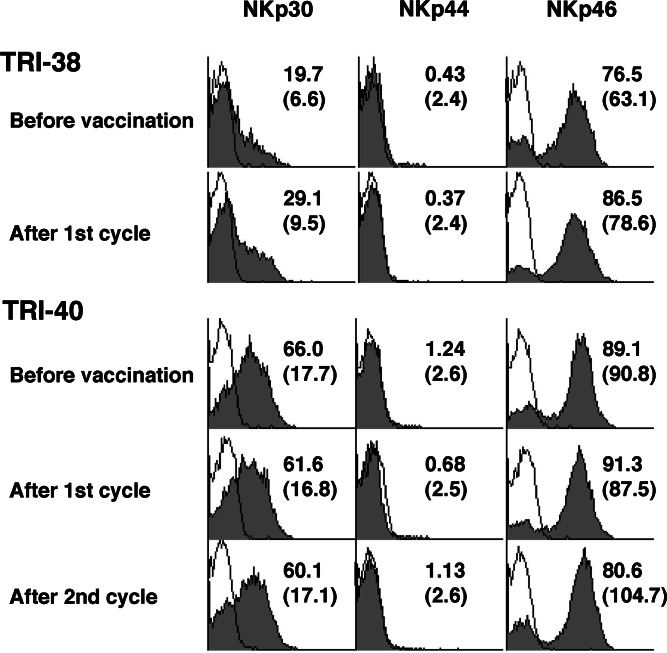
Recent reports revealed that dendritic cell (DC)-natural killer (NK) cell interaction plays an important role in tumor immunity, but few DC vaccine studies have attempted to evaluate the non-specific, yet potentially clinically relevant, NK response to immunization. In this study, we first analyzed in vitro activation of NK cells by DCs similar to those used in clinical trials. Subsequently, NK cell responses were analyzed in a phase I clinical trial of a vaccine consisting of autologous DCs loaded with a fowlpox vector encoding CEA. The data were compared with the clinical outcome of the patients. DC enhances NK activity in vitro, partly by sustaining NK cell survival and by enhancing the expression of NK-activating receptors, including NKp46 and NKG2D. Among nine patients in our clinical trial, NK cytolytic activity increased in four (range 2.5-5 times greater lytic activity) including three who had increased NK cell frequency, was stable in two and decreased in three. NKp46 and NKG2D expression showed a good correlation with the patients' NK activity. When patients were grouped by clinical activity (stable disease/no evidence of disease (stable/NE, n=5) vs progressive disease (N=4) at 3 months), the majority in the stable/NE group had increases in NK activity (P=0.016). Anti-CEA T cell response was enhanced in all the nine patients analyzed, but was not significantly different between the two groups (P=0.14). Thus, NK responses following DC vaccination may correlate more closely with clinical outcome than do T cell responses. Monitoring of NK response during vaccine studies should be routinely performed.


Figures

Fig. 1
Fig. 1
NK cell activation by DC–NK cell interaction in vitro. DCs were cultured from flask adherent cells in AIM V media containing rh-GM-CSF and rh-IL-4 for 7 days. CD56 MicroBeads (Miltenyi Biotec) was used to deplete NK cells from PBLs and DC population. One half of the DCs were infected with rF-CEA(6D)-TRICOM at moi 5 and incubated overnight. Then, PBLs or NK-depleted PBLs were incubated alone or with mock-infected (mockDC) or rF-CEA(6D)-TRICOM-infected DCs (tricomDC). a, b After a 3-day culture, the cells were harvested and analyzed for NK cell percentages, NK cell viability (a) and receptor expression on NK cells (b). Cells were stained with anti-CD3-FITC, anti-CD56-APC, 7-AAD and PE-labeled antibodies for the following antigens: CD69, CD161, CD244 (2B4), NKp30, NKp44, NKp46, NKG2A, NKG2C, and NKG2D. a Percentages of CD56+CD3- NK cells are shown in each dot plot. In each histogram, percentages of dead cells (7-AAD positive) in total NK cells are shown. b Percentages of receptor positive cells are shown in each histogram and mean fluorescence intensities are shown in parentheses. c, d After a1-week culture, the cells were harvested for the following assays. Representative data of two experiments with similar results are shown. c Culture supernatants were collected and their cytokine concentrations were analyzed by Cytometric Beads Array assay and IL-12 ELISA. d Cells were used as effector cells in standard 4 h 51Cr release assay. K562 cells were used as target cells. Open symbols PBLs cultured alone or with DCs. Filled symbols NK-depleted PBLs cultured alone or with DCs. Square symbols PBLs cultured without DC addition. Triangle symbols PBLs cultured with mock-infected DCs (mockDC). Circle symbols PBLs cultured with fp-TRICOM-infected DCs (tricomDC)
Fig. 2
Fig. 2
Changes of NK activity in patients vaccinated with rF-CEA(6D)-TRICOM-infected DCs. Frozen stocked PBMCs were thawed the day before the assay, and were incubated overnight in RPMI1640 medium+10% human AB serum with or without IL-2 (1,000 IU/ml). K562 cells labeled with 51Cr were used as target cells. Raji cells were used as negative controls. Three to five thousand K562 cells were put into each well of 96-well U-bottom plates, and PBMCs were added to make appropriate effector:target ratios. After 4 h of incubation, 51Cr release was analyzed by gamma counter. TRI-06 and TRI-32 underwent one cycle of vaccination, whereas TRI-21 and TRI-22 had two cycles. Square before vaccination, circle after first cycle of vaccination, triangle after second cycle of vaccination, filled symbol with IL-2, open symbol without IL-2
Fig. 3
Fig. 3
Changes of NK activity in PBMCs and purified NK cells. Frozen stocked PBMCs were thawed the day before the assay, and were incubated with or without IL-2 (1,000 IU/ml). NK cells were purified using NK Cell Isolation Kit after overnight incubation. K562 cells labeled with 51Cr were used as target cells. Isolated NK cells were put to 96-well plates to make 10:1 and 2:1 effector:target ratios. Square before vaccination, circle after first cycle of vaccination, triangle after second cycle of vaccination, filled symbol with IL-2, open symbol without IL-2
Fig. 4
Fig. 4
Expression of natural killing receptors on NK cells in patients vaccinated with rF-CEA(6D)-TRICOM-infected DCs. Frozen stocked PBMCs were thawed the day before the staining. Cells were stained with FITC-labeled anti-CD3, PerCP-labeled anti-CD45, APC-labeled anti-CD56 and PE-labeled anti-NKp30, 44 or 46. CD45+ FSClow SSClow CD3-CD56+ NK cells were analyzed for their expression of natural killing receptors. Two representative cases, which had increased NK activities after vaccination, are shown. Filled histograms show the NK receptor staining and solid lines show isotype controls. The numbers indicated in histogram show percentage of positive cells and the numbers in parentheses show mean fluorescence intensity
Fig. 5
Fig. 5
Expression of NK-activating receptors and inhibitory receptors on NK cells. Frozen stocked PBMCs were thawed the day before the staining. Cells were stained with FITC-labeled anti-CD3, PerCP-labeled anti-CD45, APC-labeled anti-CD56 and PE-labeled anti-CD69, 2B4 (CD244), NKG2A, NKG2C or NKG2D. CD45+ FSClow SSClow CD3-CD56+ NK cells were analyzed for their expression of these receptors. Two representative cases are shown, TRI-32: decreased NK activity, TRI-40: increased NK activity after vaccination. Filled histograms show the NK receptor staining and solid lines show isotype controls. The numbers indicated in the histogram show the percentage of positive cells and the numbers in parentheses show mean fluorescence intensity

Similar articles

Cited by

References

    1. Morse MA, Clay TM, Hobeika AC, Osada T, Khan S, Chui S, Niedzwiecki D, Panicali D, Schlom J, Lyerly HK. Phase I study of immunization with dendritic cells modified with fowlpox encoding carcinoembryonic antigen and costimulatory molecules. Clin Cancer Res. 2005;11:3017–3024. doi: 10.1158/1078-0432.CCR-04-2172. - DOI - PubMed
    1. Yokoyama WM, Kim S, French AR. The dynamic life of natural killer cells. Annu Rev Immunol. 2004;22:405–429. doi: 10.1146/annurev.immunol.22.012703.104711. - DOI - PubMed
    1. Glas R, Franksson L, Une C, Eloranta ML, Ohlen C, Orn A, Karre K. Recruitment and activation of natural killer (NK) cells in vivo determined by the target cell phenotype. An adaptive component of NK cell-mediated responses. J Exp Med. 2000;191:129–138. doi: 10.1084/jem.191.1.129. - DOI - PMC - PubMed
    1. Fernandez NC, Lozier A, Flament C, Ricciardi-Castagnoli P, Bellet D, Suter M, Perricaudet M, Tursz T, Maraskovsky E, Zitvogel L. Dendritic cells directly trigger NK cell functions: cross-talk relevant in innate anti-tumor immune responses in vivo. Nat Med. 1999;5:405–411. doi: 10.1038/7403. - DOI - PubMed
    1. Ferlazzo G, Tsang ML, Moretta L, Melioli G, Steinman RM, Munz C. Human dendritic cells activate resting natural killer (NK) cells and are recognized via the NKp30 receptor by activated NK cells. J Exp Med. 2002;195:343–351. doi: 10.1084/jem.20011149. - DOI - PMC - PubMed

Publication types

MeSH terms

Substances