. 2006 May;55(5):569-78.
doi: 10.1007/s00262-005-0037-2. Epub 2005 Nov 5.

An intracellular antibody can suppress tumorigenicity in hepatitis B virus X-expressing cells

Affiliations

An intracellular antibody can suppress tumorigenicity in hepatitis B virus X-expressing cells

Young-Hee Jin et al. Cancer Immunol Immunother. 2006 May.

Abstract

Although the hepatitis B virus X protein (HBx) is thought to play a causative role in the development of hepatocellular carcinoma, it is not yet known whether interfering with HBx function may affect the cellular transformation of HBx-expressing tumor cells. To address this question, we adopted an intracellular antibody fragment expression approach to block the function of HBx. Expression of a single-chain variable fragment (scFv) specific to HBx (designated as H7scFv) inhibited HBx-dependent cellular transactivation. Furthermore, H7scFv suppressed the growth of HBx-expressing tumor cells in both soft agar and nude mice. The suppressive effect of H7scFv on tumorigenicity appeared not to be mediated by inhibition of HBx-induced growth stimulation since the growth rate of these cells was not affected significantly by H7scFv expression. In conclusion, these data suggest that the HBx-dependent transformed phenotype is reversible and that HBx may be a good molecular target for the treatment of HBV-related tumors.


Figures

Fig. 1
Fig. 1
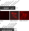
Transient expression of H7scFv and S2E1scFv in NHBx1. NHBx1 cells were transiently transfected with pCMV-Myc, pCMV-Myc-H7scFv or pCMV-Myc-S2E1scFv. a After 24 h, the lysate from transfected cells was analyzed by Western blotting with anti-Myc-Ab and HRP-conjugated antimouse IgG Ab. Expressed proteins appeared on approximately 30 kDa size band corresponding to the intracellularly expressed scFv. b One day after transfection, transiently expressed scFv proteins in NHBx1 cell was observed as bright red fluorescence signals by indirect immunofluorescence staining with anti-Myc Ab and cyanine 3-conjugated rabbit affinity purified Ab to mouse IgG using Confocal microscopy. The expressed scFvs were located in the cytoplasm
Fig. 2
Fig. 2
Association of H7scFv with HBx in NIH3T3 cells. NIH3T3 cells were cotransfected with pIRES2/Flag-HBx and either one of pCMV/Myc-H7scFv or pCMV/Myc–S2E1scFv. After 24 h incubation, cells were treated with or without 10μM clasto-lactacystin β–lactone, an irreversible proteasome inhibitor. Two hours later, cells were lysed and lysates were incubated with the anti-Flag Ab. The immune complex was precipitated with protein G plus protein A agarose and analyzed by SDS-PAGE, followed by immunodetection of HBx with anti-Flag Ab. Transfection with pIRES2/Flag-HBx alone served as controls (upper panel). Immunodetection of scFv with anti-Myc Ab was performed in the same membrane (lower panel)
Fig. 3
Fig. 3
Expression of scFv and HBx in stable cell lines. a H7scFv and S2E1scFv transcripts were analyzed by RT-PCR using primers to the full fragment of scFv in selected stable cell lines. pCMV/Myc-H7scFv and pCMV/Myc-S2E1scFv were used as a positive control. b Expression of H7scFv and S2E1scFv in stable cell lines was observed by indirect immunofluorescence staining with anti-Myc Ab and cyanine 3-conjugated rabbit affinity purified Ab to mouse IgG. H7scFv and S2E1scFv proteins were detected as bright red fluorescent signals. c HBx transcript in selected stable transfectants was analyzed by RT-PCR using primers to HBx. pCMV/HA-HBx was used as a positive control
Fig. 4
Fig. 4
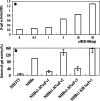
The effect of H7scFv expression on the transactivation of HBx in stable cell lines. a To confirm the transactivation ability of HBx on the SV40 early promoter, β-galactosidase (β-gal) activity was measured in NIH3T3 cells transfected with the indicated amount of pCMV-HA/HBx and pCH110 which contains the β-galactosidase gene under the control of SV40 ori/early promoter. The β-galactosidase activity was measured by color change in the lysate of transfected NIH3T3 cells as described in Materials and methods. β-galactosidase activity was increased in a HBx level-dependent manner. The experiments were repeated three times and similar trends were observed in each case. b To analyze the transactivation of HBx in stable cell lines, β-galactosidase activity was measured in the lysate of stable cell lines transfected with pCH110. The lysates of NHBx1-H7scFv1 and NHBx1-H7scFv3 cell lines caused a reduction in β-galactosidase activity as compared to the lysate of parental NHBx1 cells and NHBx1 cells expressing S2E1scFv. Relative β-gal activity (OD of each lysate/OD of NHBx1 lysate×100) was calculated. The mean percentage was obtained through three independent experiments in duplicate. Error bars represent standard error
Fig. 5
Fig. 5
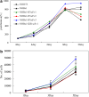
Proliferation of NHBx1-H7scFv stable cell lines. a To analyze the growth of NHBx1-H7scFv1–3 and NHBx1-S2E1scFv1, 3×10² cells were inoculated onto 96-well culture plates. The proliferation rate was analyzed by MTT assay on the 2nd, 4th, 6th, 8th and 10th days after inoculation. The experiments were repeated three times and similar trends were observed in each case. b Onto 96-well culture plates 1×10³ cells were inoculated. The number of cells was analyzed by MTT assay on the 1st, 3rd and 5th days after inoculation. Standard curves by cell number were devised as described in Materials and methods. The mean cell numbers of three independent experiments in triplicate are shown. Error bars represent standard error
Fig. 6
Fig. 6
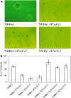
The effect of H7scFv expression on the growth of stable cell lines in soft agar. Stable cell lines (1×10⁴ cells/ml) were plated into soft agar, as described in Materials and methods. On day 21, colonies were photographed a and the number of colony greater than 0.2 mm was counted. b The mean numbers of three independent experiments, performed in duplicate, are shown. Error bars represent standard error
Fig. 7
Fig. 7
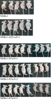
Tumorigenicity of NHBx1-H7scFv stable cell lines in nude mice. Cells (2×10⁶) of each stable cell lines were injected into nude mice and the mice were photographed 8 weeks later

Similar articles

Cited by

References

    1. Unsal H, Yakicier C, Marcais C, Kew M, Volkmann M, Zentgraf H, Isselbacher KJ, Ozturk M. Genetic heterogeneity of hepatocellular carcinoma. Proc Natl Acad Sci USA. 1994;91:822–826. doi: 10.1073/pnas.91.2.822. - DOI - PMC - PubMed
    1. Diamantis ID, McGandy CE, Chen TJ, Liaw YF, Gudat F, Bianchi L. Hepatitis B X-gene expression in hepatocellular carcinoma. J Hepatol. 1992;15:400–403. doi: 10.1016/0168-8278(92)90077-3. - DOI - PubMed
    1. Shirakata Y, Kawada M, Fujiki Y, Sano H, Oda M, Yaginuma K, Kobayashi M, Koike K. The X gene of hepatitis B virus induced growth stimulation and tumorigenic transformation of mouse NIH3T3 cells. Jpn J Cancer Res. 1989;80:617–621. - PMC - PubMed
    1. Kim CM, Koike K, Saito I, Miyamura T, Jay G. HBx gene of hepatitis B virus induces liver cancer in transgenic mice. Nature. 1991;351:317–320. doi: 10.1038/351317a0. - DOI - PubMed
    1. Koike K, Moriya K, Iino S, Yotsuyanagi H, Endo Y, Miyamura T, Kurokawa K. High-level expression of hepatitis B virus HBx gene and hepatocarcinogenesis in transgenic mice. Hepatology. 1994;19:810–819. doi: 10.1002/hep.1840190403. - DOI - PubMed

Publication types

MeSH terms

Substances

LinkOut - more resources