. 2006 Feb;235(2):315-26.
doi: 10.1002/dvdy.20626.

Discrete pulses of molting hormone, 20-hydroxyecdysone, during late larval development of Drosophila melanogaster: correlations with changes in gene activity

Affiliations

Discrete pulses of molting hormone, 20-hydroxyecdysone, during late larval development of Drosophila melanogaster: correlations with changes in gene activity

James T Warren et al. Dev Dyn. 2006 Feb.

Abstract

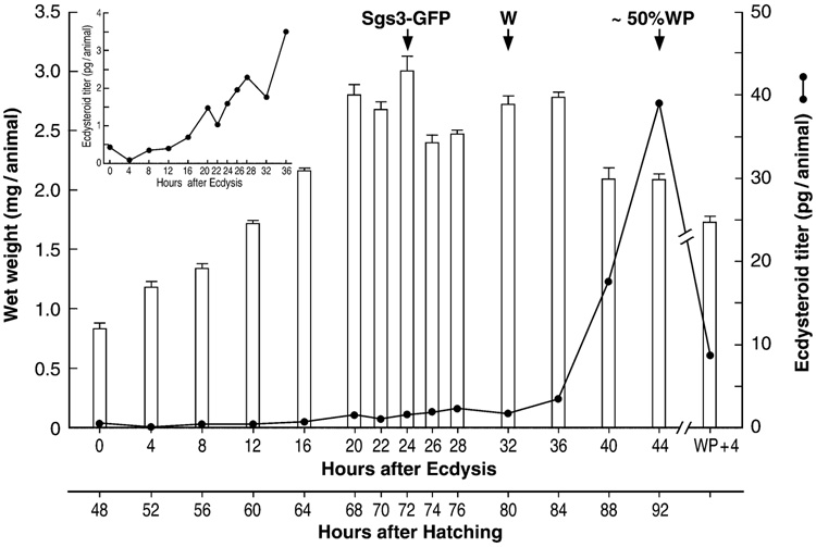
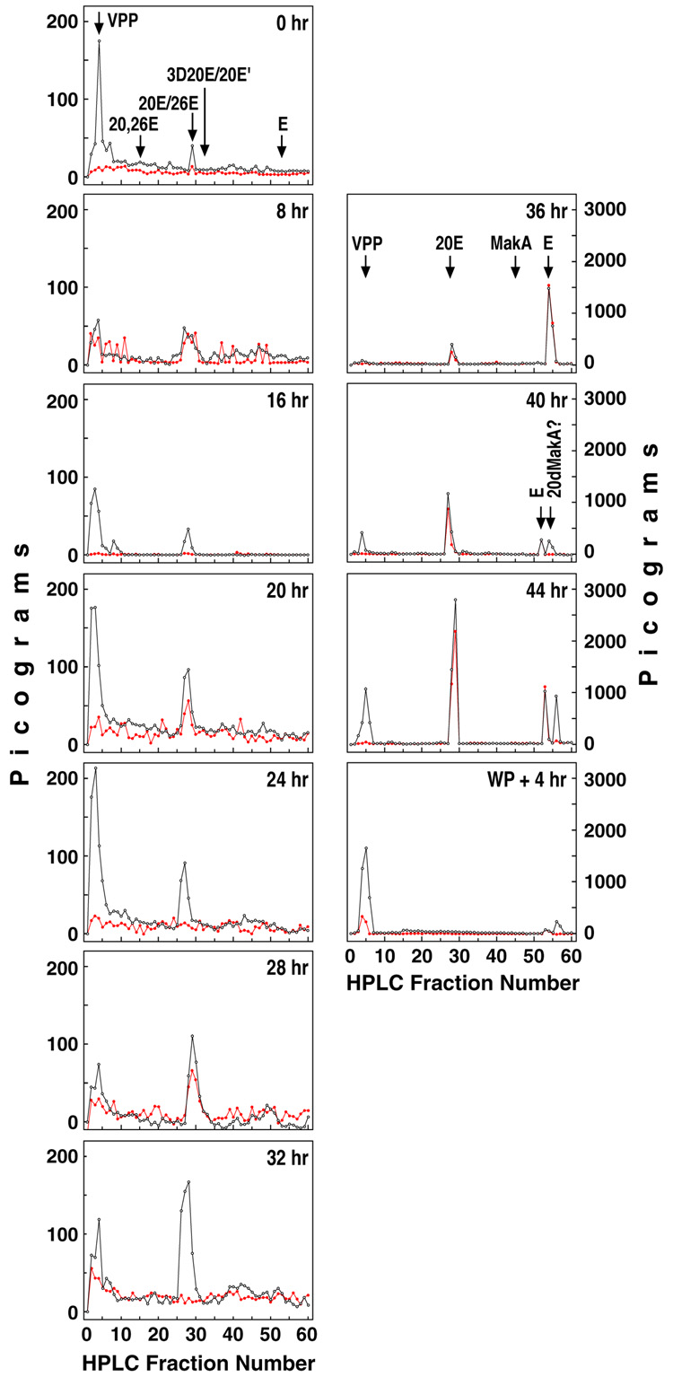
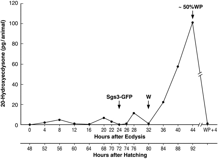
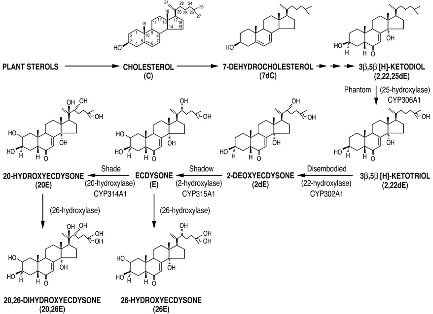
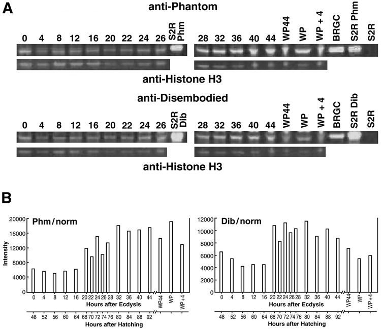
Periodic pulses of the insect steroid molting hormone 20-hydroxyecdysone (20E), acting via its nuclear receptor complex (EcR/USP), control gene expression at many stages throughout Drosophila development. However, during the last larval instar of some lepidopteran insects, subtle changes in titers of ecdysteroids have been documented, including the so-called "commitment peak." This small elevation of 20E reprograms the larva for metamorphosis to the pupa. Similar periods of ecdysteroid immunoreactivity have been observed during the last larval instar of Drosophila. However, due to low amplitude and short duration, along with small body size and staging difficulties, their timing and ecdysteroid composition have remained uncertain. Employing a rigorous regimen of Drosophila culture and a salivary gland reporter gene, Sgs3-GFP, we used RP-HPLC and differential ecdysteroid RIA analysis to determine whole body titers of 20E during the last larval instar. Three small peaks of 20E were observed at 8, 20, and 28 hr following ecdysis, prior to the well-characterized large peak around the time of pupariation. The possible regulation of 20E levels by biosynthetic P450 enzymes and the roles of these early peaks in coordinating gene expression and late larval development are discussed.


Figures

Figure 1
Figure 1
Larval growth curve and crude ecdysteroid titer. Shown are the mean wet weight (mg/animal +/− SE) and the whole body ecdysteroid titer in pg ecdysteroid/animal, as measured using the SHO3 antiserum, during the third larval instar. Sgs3-GFP denotes the time that the Sgs3-GFP fusion protein becomes significantly expressed in the larval salivary glands. (W), beginning of wandering; (~50% WP), about 50% of animals had formed white puparia (WP) by 44 hr after ecdysis. Their titer was identical to that of the larvae at 44 hr. (WP + 4), 4 hr after formation of a white puparia. Inset was included to clarify the small multiple titer elevations and peaks occurring at 8, 20 and 28 hr.
Figure 2
Figure 2
RP-HPLC/RIA analysis of whole body extracts during the third larval instar in Drosophila. The ecdysteroid content, as measured by RIA using either the H22 (black) or SHO3 antisera (red) is shown for each HPLC fraction (1 ml) in selected samples as indicated (in hours after ecdysis to the third instar). (VPP), very polar products; (E), ecdysone; (20E), 20-hydroxyecdysone; (26E), 26-hydroxyecdysone; (20,26E), 20,26-dihydroxyecdysone; (3D20E), 3-dehydro-20-hydroxyecdysone; (20E'), 3-α-epi-20-hydroxyecdysone; (MakA), makisterone A; (20dMakA), 20-deoxymakisterone A (24-methylecdysone); 44, combined larval and white puparial (44WP) extracts; (WP + 4 hr), pre-pupae 4 hr after formation of white puparia.
Figure 3
Figure 3
Whole body titer of 20-hydroxyecdysone during the third larval instar in Drosophila. The concentration of 20E was determined using RIA with the SHO3 antisera following sample resolution by RP-HPLC. A correction for 20E cross-reactivity was made, i.e. a 5-fold increase relative to E. Sgs3-GFP denotes the time that the Sgs3-GFP fusion protein is first significantly expressed in the larval salivary glands. (W), beginning of wandering; (~50% WP), about 50% of animals had formed white puparia (WP) by 44 hr after ecdysis. Extracts of larvae and pupae at 44 hr were combined for this analysis. (WP + 4), pre-pupae 4 hr after formation of white puparia.
Figure 4
Figure 4
Biosynthesis and metabolism of E and 20E. Four cytochrome P450 (CYP) enzymes have thus far been identified as catalyzing the last four hydroxylation reactions in Drosophila ecdysteroidogenesis. Also shown are 26E and 20,26E, the products of an inactivation reaction, i.e. ecdysteroid 26-hydroxylation, catalyzed by an as yet uncharacterized CYP enzyme.
Figure 5
Figure 5
Profiles of Dib and Phm protein expression. Protein samples where prepared as described in Methods, separated on 4–12% Bis-Tris NuPAGE gels and blotted onto PDVF membranes. (A). Shown are representative blots probed for the biosynthetic enzymes Phm (top set, anti-Phantom) or Dib (bottom set, anti-Disembodied). Below each are shown the same blots probed for the protein loading control histone H3 (tubulin E7 not shown). Times for larvae are in hours after ecdysis to the third instar. WP44, animals at 44 hr that had developed into white puparia; (WP), additional samples of white puparia; WP + 4, pre-pupae 4 hrs after white puparium formation. (B). Protein levels where quantified using Odyssey software (Licor). The average of two measurements each for tubulin E7 and histone H3 were used to derive a normalized average value for total protein loaded per lane. Two measurements each for Phm and Dib were used to give an average amount for each. This average Phm or Dib value was divided by the normalized averaged value for total protein to yield the intensity numbers shown in the graphs (y-axis). Two blots where run for each sample and an average value was obtained for each time point. BRGC, brain ring gland complex; S2R, protein extracted from transfected S2R cells expressing either Phm or Dib or else an un-transfected S2R control.

References

    1. Andres AJ, Fletcher JC, Karim FD, Thummel CS. Molecular analysis of the initiation of insect metamorphosis: a comparative study of Drosophila ecdysteroid-regulated transcription. Dev Biol. 1993;160:388–404. - PubMed
    1. Andres AJ, Thummel CS. Methods for quantitative analysis of transcription in larvae and prepupae. In: Goldstein LSB, Fyrberg EA, editors. Drosophila melanogaster: Practical uses in Cell and Molecular Biology. San Diego: Academic Press; 1994. pp. 565–573. - PubMed
    1. Ashburner M. Drosophila: A laboratory handbook. Cold Spring Harbor: Cold Spring Harbor Laboratory Press; 1989. Developmental biology; pp. 139–298.
    1. Ashburner M, Chihara C, Meltzer P, Richards G. Temporal control of puffing activity in polytene chromosomes. Cold Spring Harbor Symp. Quant Biol. 1974;38:655–662. - PubMed
    1. Belinski-Deutsch S, Busson D, Lamour-Audit C, Porcheron P, Moriniere M, Berreur P. Relations between ecdysteroid levels and pupal development in the ecd-1 temperature-sensitive mutant of Drosophila melanogaster. J Insect Physiol. 1983;29:509–514.

Publication types

MeSH terms

LinkOut - more resources