. 2005 Nov 7;171(3):505-16.
doi: 10.1083/jcb.200504124.

FAK signaling is critical for ErbB-2/ErbB-3 receptor cooperation for oncogenic transformation and invasion

Affiliations

FAK signaling is critical for ErbB-2/ErbB-3 receptor cooperation for oncogenic transformation and invasion

Naciba Benlimame et al. J Cell Biol. .

Abstract

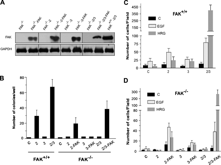
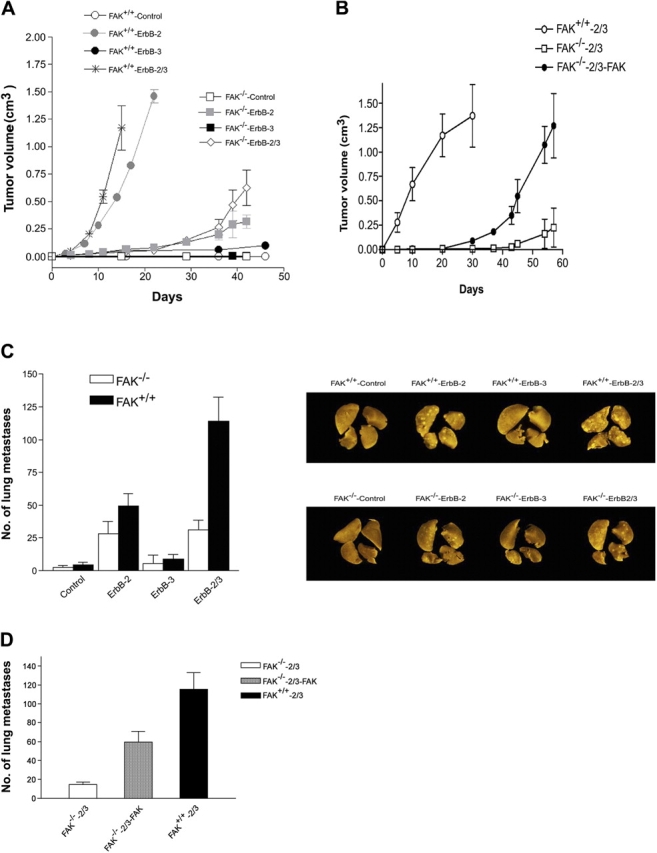
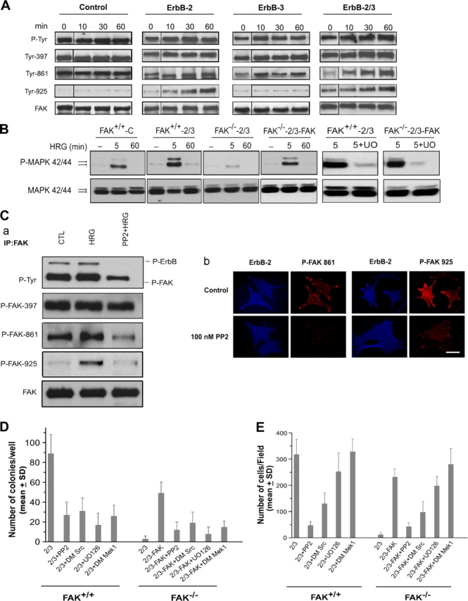
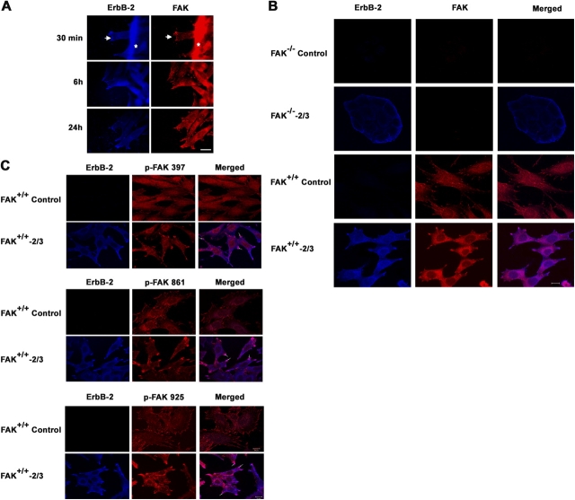
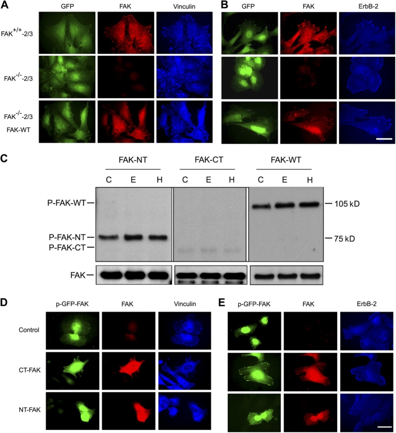
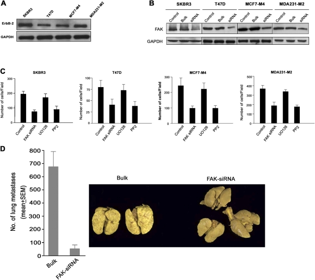
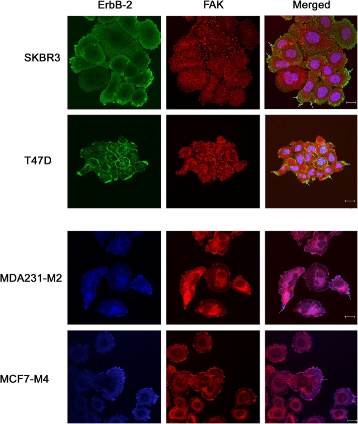
The overexpression of members of the ErbB tyrosine kinase receptor family has been associated with cancer progression. We demonstrate that focal adhesion kinase (FAK) is essential for oncogenic transformation and cell invasion that is induced by ErbB-2 and -3 receptor signaling. ErbB-2/3 overexpression in FAK-deficient cells fails to promote cell transformation and rescue chemotaxis deficiency. Restoration of FAK rescues both oncogenic transformation and invasion that is induced by ErbB-2/3 in vitro and in vivo. In contrast, the inhibition of FAK in FAK-proficient invasive cancer cells prevented cell invasion and metastasis formation. The activation of ErbB-2/3 regulates FAK phosphorylation at Tyr-397, -861, and -925. ErbB-induced oncogenic transformation correlates with the ability of FAK to restore ErbB-2/3-induced mitogen-activated protein kinase (MAPK) activation; the inhibition of MAPK prevented oncogenic transformation. In contrast, the inhibition of Src but not MAPK prevented ErbB-FAK-induced chemotaxis. In migratory cells, activated ErbB-2/3 receptors colocalize with activated FAK at cell protrusions. This colocalization requires intact FAK. In summary, distinct FAK signaling has an essential function in ErbB-induced oncogenesis and invasiveness.


Figures

Figure 1.
Figure 1.
Overexpression of ErbB-2, -3, and -2/3 in FAK + / + and FAK / cells. (A) Cells expressing control retroviral particles or ErbB receptors were subjected to Western blotting using specific ErbB antibodies as described in Materials and methods. (B) Cells were fixed and double stained for ErbB-2 and -3 using specific antibodies and were examined by immunofluorescence microscopy. Note that FAK−/− and FAK+/+ control cells do not express any ErbB-2 or -3 receptors, whereas FAK−/−-2/3 and FAK+/+-2/3 exhibited strong labeling for both receptors. Bar, 40 μm. (C) Cells were serum starved for 24 h and kept unstimulated (control, C) or were stimulated with 20 ng/ml EGF (E) or HRG (H) for 10 min. Cell lysates were immunoprecipitated with anti-ErbB, and the blots were probed using antiphosphotyrosine antibody and reprobed with the corresponding ErbB-specific antibody.
Figure 2.
Figure 2.
FAK is essential for ErbB-induced oncogenic transformation and chemotaxis. (A) Western blot analysis to show FAK expression status in FAK−/− cells expressing various ErbB receptor combinations and their matched cells in which wild-type FAK was reconstituted by stable transfection, as indicated in Materials and methods. FAK+/+–ErbB-2/3 cells are included as a control. (B) Oncogenic property of FAK−/−- and FAK+/+-expressing ErbB-2/3 receptors. Cells were cultured in medium containing soft agarose, and colony formation was determined 4 wk later by counting the number of cell foci of >20 μm in diameter (Fig. S2 A, available at http://www.jcb.org/cgi/content/full/jcb.200504124/DC1). Each bar on the graph represents the mean number of colonies per well from three independent experiments ± SD. (C and D) ErbB-induced cell invasion in FAK+/+ (C), FAK−/−, and FAK−/− cells in which FAK was restored (D). FAK+/+, FAK−/−, and FAK−/−-reconstituted cells expressing ErbB receptors were cultured in the upper chamber, whereas EGF or HRG was used as a chemoattractant in the lower chamber. Each bar of the graphs represents the mean ± SD (error bars) of invading cells from three independent experiments. C, control.
Figure 3.
Figure 3.
FAK is required for ErbB-induced tumor progression and tumor invasion in vivo, whereas the restoration of FAK in FAK / cells rescued the deficiency in tumor invasion. (A) Tumor growth kinetics after subcutaneous implantation of cells into the flank of Scid mice. (B) Tumor growth kinetics after subcutaneous implantation of FAK−/−-2/3 and FAK−/−-2/3–reconstituted cells (FAK−/−-2/3–FAK); FAK+/+-2/3 cells were used as positive controls. Tumor growth was monitored over time as indicated in Materials and methods. Each point represents the mean of five to eight mice ± SEM. (C) Quantification of lung surface metastases induced by intravenous cell administration. (a) Mean surface lung metastases (n = 8–10) ± SEM. (b) Representative lungs from mice inoculated with FAK−/− and FAK+/+ control cells (expressing empty retroviral particles) and cells overexpressing ErbB-2, -3, or -2/3 receptors. (D) Mean lung metastases (n = 8) ± SEM (error bars) induced by FAK−/−-2/3 or FAK-reconstituted FAK−/−-2/3 (FAK−/−-2/3–FAK). FAK+/+-2/3 cells were used as positive controls.
Figure 4.
Figure 4.
ErbB-induced oncogenic transformation and invasion are mediated via distinct FAK signaling. (A) Differential regulation of FAK phosphorylation by ErbB-2 and -3 receptors. Serum-starved cells were stimulated with 20 ng/ml EGF (control cells) or HRG (ErbB-2, -3, and -2/3–overexpressing cells) for the indicated times. Proteins were immunoprecipitated with anti-FAK and probed with antiphosphotyrosine antibody, antiphospho-FAK antibodies specific to different residues, and anti-FAK antibody. (B) Differential regulation of MAPK phosphorylation by ErbB-2 and -3. Cell lysates from HRG-stimulated adherent cells were blotted with P-MAPK and reprobed with MAPK antibody as indicated in Materials and methods. The figure shows an increase in P-MAPK after 5 min of stimulation with HRG in FAK+/+-2/3 cells and FAK-reconstituted FAK−/−-2/3 cells compared with control cells. Exposure to UO126 strongly inhibited P-MAPK in FAK-proficient cells. (C) Inhibition of Src reduced HRG-induced FAK phosphorylation at Tyr-861 and -925. FAK+/+-2/3 cells were serum starved for 24 h and pretreated with PP2 at 100 nM for 60 min followed by treatment with 20 ng/ml HRG for 30 min. (a) Whole cell lysates were immunoprecipitated with anti-FAK and probed with antiphosphotyrosine antibody. Membranes were subsequently reprobed with antiphospho-FAK antibodies specific to different residues and total FAK. (b) FAK+/+-2/3 cells were coimmunostained for ErbB-2 and the indicated phospho-FAK in the absence or presence of PP2. Bar, 40 μm. (D) Inhibition of colony formation on agar by Src and MAPK inhibition. Cells were cultured in medium containing soft agarose either in the absence and presence of PP2 or UO126 or were transfected with dominant mutants for MEK1 or Src. Colony formation was determined 4 wk later by counting the number of cell foci. (E) Inhibition of cell invasion by Src but not MAPK inhibition. Cells were cultured in the upper chamber of the Boyden chamber in the absence and presence of PP2 or UO126 or after being transfected with dominant mutants for MEK1 or Src. HRG was used as a chemoattractant in the lower chamber. Each bar of the graphs represents the mean ± SD (error bars) of invading cells from three independent experiments.
Figure 5.
Figure 5.
ErbB-2 colocalizes with FAK at focal adhesions in motile cells. (A) ErbB-2 and FAK colocalize in motile FAK+/+-2/3 cells. Confluent cells were scratch wounded and allowed to heal for the indicated time points before fixation. Cells were then coimmunostained with anti–ErbB-2 and anti-FAK antibodies followed by appropriate secondary antibodies as described in Materials and methods. Note that both ErbB-2 and FAK are recruited into newly formed lamellipodia near the leading edge of the wounded cells during cell migration to the acellular area (30 min). Typical ventral focal contacts that were stained for FAK become detectable 6 h after wound healing and become more pronounced after 24 h. Arrowheads indicate the newly formed protrusions. Stars indicate the folded cell layer at the wounded area. Bar, 30 μm. (B) Colocalization of ErbB-2 and FAK at the cell protrusion. Cells were fixed, permeabilized, and double immunostained with anti–ErbB-2 and anti-FAK antibodies followed by appropriate secondary antibodies conjugated either to aminomethylcoumarin (AMCA) or Texas red to detect ErbB-2 and FAK, respectively. The figure shows that FAK−/− control cells were negatively stained for both ErbB-2 and FAK, whereas FAK−/−-2/3 cells were strongly labeled for ErbB-2 receptors, which were homogeneously distributed around the cell periphery. In contrast, FAK+/+ control cells exhibit strong labeling of FAK, which was localized to cell extensions and ventral focal contact sites within the cells, whereas FAK+/+-2/3 cells exhibit strong labeling of FAK at cell protrusions, which colocalize partially with ErbB-2 receptors as revealed by dual-color merged confocal images. (C) Tyrosine-phosphorylated FAK colocalized with ErbB-2. FAK+/+ control cells and FAK+/+-2/3 were grown in complete medium, fixed, and immunostained with ErbB-2 in combination with either FAK Tyr-397, -861, or -925 antibodies. Confocal microscopy reveals partial colocalization of phospho-FAK with ErbB-2 receptor at the cell protrusions (arrows). In contrast, control cells that do not express any ErbB exhibit homogeneous distribution of phospho-FAK at the focal contacts. (B and C) Bars, 50 μm.
Figure 6.
Figure 6.
Restoration of FAK in FAK / cells rescued focal adhesions and ErbB-2 relocalization to focal protrusions. (A) FAK−/− cells cooverexpressing ErbB-2 and -3 receptors were stably transfected with wild-type FAK as described in Materials and methods. Double immunofluorescence labeling of FAK and vinculin, a marker for focal adhesion, demonstrated that FAK−/−-2/3 cells in which FAK was restored (right) exhibited many focal adhesions that were labeled for both FAK and vinculin, which is similar to normal FAK+/+-2/3 cells. (B) Double immunofluorescence of FAK and ErbB-2 revealed that the restoration of wild-type FAK in FAK−/−-2/3 cells induces the relocalization of ErbB-2 receptors from the cell membrane to fingerlike protrusions. This ErbB-2 pattern is similar to FAK+/+-2/3 cells in which FAK and ErbB-2 receptors colocalized at cell protrusions. Bar, 30 μm. (C) Expression of NH2 terminus (NT) and COOH terminus (CT) FAK in FAK−/−-2/3. Cells expressing the NT or CT were stimulated without (C) or with 20 ng/ml EGF (E) or 20 ng/ml HRG (H) for 30 min. Equal amounts of protein were immunoprecipitated with anti–FAK-A17, which targets at NT-FAK, or anti-FAK–C-20 which targets at CT-FAK. Blots were probed with antiphosphotyrosine antibody. (D and E) Immunofluorescence analysis. FAK−/−-2/3 cells expressing control, CT-FAK–GFP, or NT-FAK–GFP fusion protein (see Materials and methods). After transfection, the cells were incubated in complete medium for 24 h and were fixed and coimmunostained for FAK and vinculin (D) or FAK and ErbB-2 receptor (E). Note that focal adhesions were seen only when cells were transfected with CT-FAK but not with NT-FAK (D). In contrast, cells transfected with CT-FAK exhibited a homogeneous distribution of ErbB-2 receptors at the cell membrane, whereas transfection with NT-FAK exhibited a reduced staining of ErbB-2 throughout the cytoplasm (E). Bar, 30 μm.
Figure 7.
Figure 7.
FAK regulates ErbB-induced cell invasion and metastasis formation that is induced by human breast cancer cells. (A) ErbB-2 expression status examined by Western blot analysis of cell extracts from cells constitutively overexpressing ErbB-2 (SKBR-3 and T47D) and metastatic cell variants (MCF7-M4 and MDA-231-M2) that were isolated in vivo from MCF7 and MDA-231 cells engineered to overexpress ErbB-2. (B) Western blots on cells stably expressing control (bulk) or FAK siRNA. Note that FAK-specific siRNA induced a marked decrease of FAK expression. GAPDH used as an internal control was unaffected. (C) Inhibition of FAK by siRNA reduced cell invasion to a similar level as observed after Src inhibition but not MAPK inhibition. Cells expressing siRNA or treated with PP2 or UO126 were cultured in the upper chamber, whereas HRG was used as a chemoattractant in the lower chamber. Each bar of the graphs represents the mean ± SD of invading cells from three independent experiments. (D) The metastatic MDA-231-M2 cells were implanted into the mammary fat pad. Animals were sacrificed 50 d later, lungs were fixed in Bouin, and surface lung metastases were counted. Each bar represents the mean of five mice ± SEM (error bars). Image is of representative lungs from these experiments.
Figure 8.
Figure 8.
ErbB-2 colocalizes with FAK at focal adhesions in invasive breast cancer cells. Cells were fixed, permeabilized, and subjected to confocal microscopy after double immunostaining with anti–ErbB-2 and anti-FAK antibodies followed by appropriate secondary antibodies conjugated either to Cy2 or Texas red (left) or conjugated to AMCA and Texas red (middle) to detect ErbB-2 and FAK, respectively. The panels show a strong labeling of FAK, which was localized to cell extensions and ventral focal contact sites within the cells. For all cell lines, dual-color merged confocal images reveal a partial colocalization of ErbB-2 with FAK (arrows). Bars, 50 μm.

References

    1. Alaoui-Jamali, M.A., D.J. Song, N. Benlimame, L. Yen, X. Deng, M. Hernandez-Perez, and T. Wang. 2003. Regulation of multiple tumor microenvironment markers by overexpression of single or paired combinations of ErbB receptors. Cancer Res. 63:3764–3774. - PubMed
    1. Alimandi, M., A. Romano, M.C. Curia, R. Muraro, P. Fedi, S.A. Aaronson, P.P. Di Fiori, and M.H. Kraus. 1995. Cooperative signaling of ErbB3 and ErbB2 in neoplastic transformation and human mammary carcinomas. Oncogene. 10:1813–1821. - PubMed
    1. Baulida, J., M.H. Kraus, M. Alimandi, P.P. Di Fiore, and G. Carpenter. 1996. All ErbB receptors other than the epidermal growth factor receptor are endocytosis impaired. J. Biol. Chem. 271:5251–5257. - PubMed
    1. Brunton, V.G., B.W. Ozanne, C. Paraskeva, and M.C. Frame. 1997. A role for epidermal growth factor receptor, c-Src and focal adhesion kinase in an in vitro model for the progression of colon cancer. Oncogene. 14:283–293. - PubMed
    1. Carragher, N.O., M.A. Westhoff, D. Riley, D.A. Potter, P. Dutt, J.S. Elce, P.A. Greer, and M.C. Frame. 2002. v-Src-induced modulation of the calpain-calpastatin proteolytic system regulates transformation. Mol. Cell. Biol. 22:257–269. - PMC - PubMed

Publication types

MeSH terms