. 2005 Nov 7;202(9):1213-23.
doi: 10.1084/jem.20051135.

TSLP-activated dendritic cells induce an inflammatory T helper type 2 cell response through OX40 ligand

Affiliations

TSLP-activated dendritic cells induce an inflammatory T helper type 2 cell response through OX40 ligand

Tomoki Ito et al. J Exp Med. .

Abstract

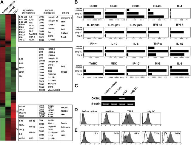
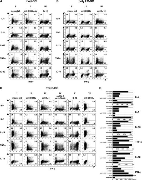
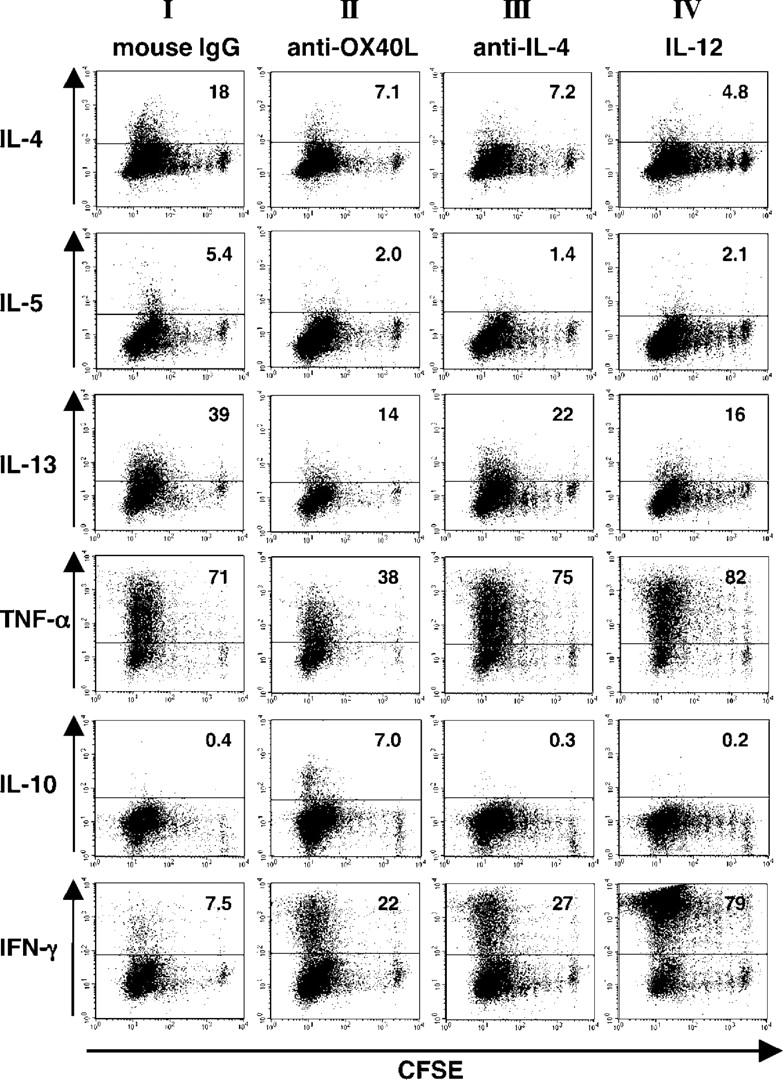
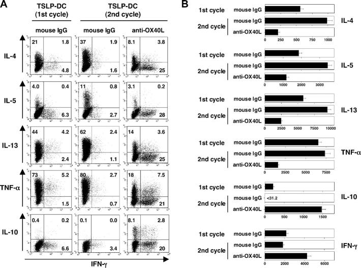
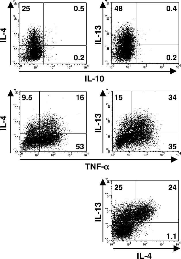
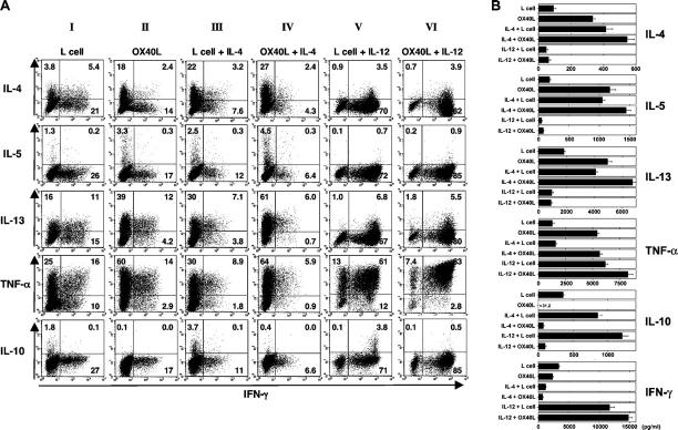
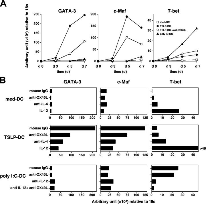
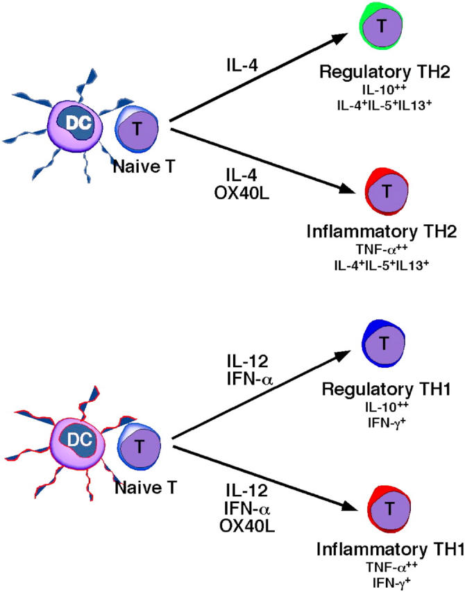
We recently showed that dendritic cells (DCs) activated by thymic stromal lymphopoietin (TSLP) prime naive CD4(+) T cells to differentiate into T helper type 2 (Th2) cells that produced high amounts of tumor necrosis factor-alpha (TNF-alpha), but no interleukin (IL)-10. Here we report that TSLP induced human DCs to express OX40 ligand (OX40L) but not IL-12. TSLP-induced OX40L on DCs was required for triggering naive CD4(+) T cells to produce IL-4, -5, and -13. We further revealed the following three novel functional properties of OX40L: (a) OX40L selectively promoted TNF-alpha, but inhibited IL-10 production in developing Th2 cells; (b) OX40L lost the ability to polarize Th2 cells in the presence of IL-12; and (c) OX40L exacerbated IL-12-induced Th1 cell inflammation by promoting TNF-alpha, while inhibiting IL-10. We conclude that OX40L on TSLP-activated DCs triggers Th2 cell polarization in the absence of IL-12, and propose that OX40L can switch IL-10-producing regulatory Th cell responses into TNF-alpha-producing inflammatory Th cell responses.


Figures

Figure 1.
Figure 1.
TSLP-DCs express OX40L. (A) Each column represents data from human blood CD11c+ immature mDCs either resting or activated by poly I:C or TSLP. The selected 2,166 genes were grouped, based on similarity in expression patterns, by hierarchical clustering as described in Materials and methods. Each row represents relative hybridization intensities of a particular gene across different samples. Colors reflect the magnitude of relative expression of a particular gene across samples. Cluster I, II, and III include genes highly expressed in poly I:C–activated DCs, resting DCs, and TSLP-activated DCs, respectively. Cluster IV includes genes highly expressed in both poly I:C–activated and TSLP-activated DCs. Under culture with different stimuli, expression profiles of the indicated genes (B) and OX40L mRNA expression (C) in DCs were analyzed at the 24-h time point by microarray and RT-PCR, respectively, and surface expression of OX40L on DCs were analyzed at the 48-h time point by flow cytometry (D). OX40L expression on TSLP-DCs were monitored at different time points (E). The staining profile of anti-OX40L mAb and isotype-matched control are shown by the shaded and open areas, respectively. The results of the gene expression profiles (B) are shown as the relative hybridization intensity level by microarray analysis. Accession number of each microarray dataset are available at http://www.ncbi.nih.gov/entrez/query.fcgi?db=gene.
Figure 2.
Figure 2.
TSLP-DC-mediated inflammatory Th2 cell response requires OX40L as a positive Th2 cell–polarizing signal and default of a lack of IL-12. CD4+ naive T cells were cocultured with med-DCs (A), poly I:C-DCs (B), or TSLP-DCs (C and D) for 7 d in the presence of the indicated neutralizing Abs or recombinant IL-12. Cytokine production by T cells was analyzed intracellularly by flow cytometry (A–C) and measured in supernatants after restimulation with anti-CD3 and anti-CD28 mAbs for 24 h by ELISA (D). Percentages of the respective cytokine-producing T cells are indicated in each dot blot profile in A–C. Error bars in D represent standard deviations of triplicate cultures. Data are from one of four independent experiments.
Figure 3.
Figure 3.
Relationship between cytokine production and cell division in T cells primed with TSLP-DCs. CFSE-labeled CD4+ naive T cells were primed with TSLP-DCs in the presence of an anti-OX40L mAb, an anti–IL-4 mAb, or recombinant IL-12 for 7 d, and then intracellular cytokines were analyzed by flow cytometry. Numbers in each dot blot profile indicate the percentages of the respective cytokine-producing T cells. Data are from one of three independent experiments.
Figure 4.
Figure 4.
TSLP-DCs further promote the generation of Th2 cells without inducing IL-10 production after two rounds of stimulation. CD4+ naive T cells were stimulated by TSLP-DCs for 7 d (one cycle of stimulation), and then these differentiated CD4+ T cells were restimulated by TSLP-DCs from the same allogeneic donor for further 7 d (two cycles of stimulation) in the presence or absence of anti-OX40L mAbs. Cytokine production by these two types of CD4+ T cells was analyzed intracellularly by flow cytometry (A) and measured in supernatants after restimulation with anti-CD3 and anti-CD28 mAbs for 24 h by ELISA (B). Percentages of the respective cytokine-producing T cells are indicated in each dot blot profile in A. Error bars in B represent standard deviations of triplicate cultures. Data are from one of three independent experiments.
Figure 5.
Figure 5.
Th2 cells induced by TSLP-DCs coexpress TNF-α but not IL-10, as analyzed by three-color intracellular cytokine staining. Naive CD4+ T cells were primed with TSLP-DCs for 7 d, and then cytokine production by the CD4+ T cells was analyzed intracellularly by flow cytometry using three patterns of triple-color staining as follows: (a) FITC-labeled anti–TNF-α + PE-labeled anti–IL-4 + APC-labeled anti–IL-10; (b) FITC-labeled anti–TNF-α + PE-labeled anti–IL-13 + APC-labeled anti–IL-10; and (c) FITC-labeled anti–TNF-α + PE-labeled anti–IL-13 + Alexa Fluor 647-labeled anti–IL-4. Percentages of the respective cytokine-producing T cells are indicated in each dot blot profile. Data are from one of three independent experiments.
Figure 6.
Figure 6.
OX40L-transfected L cells enhance TNF-α production and inhibit IL-10 production in T cells. CD4+ naive T cells were cultured with anti-CD3 and anti-CD28 mAbs on OX40L-transfected L cells and/or the indicated recombinant cytokines for 7 d. Cytokine production by CD4+ T cells was analyzed intracellularly by flow cytometry (A) and measured in supernatants after restimulation with anti-CD3 and anti-CD28 mAbs for 24 h by ELISA (B). Percentages of the respective cytokine-producing T cells are indicated in each dot blot profile in A. Error bars in B represent standard deviations of triplicate cultures. Data are from one of four independent experiments.
Figure 7.
Figure 7.
OX40L expressed by TSLP-DCs induces the expression of GATA-3 and c-Maf in T cells. Expression levels of transcriptional factors involved in Th1 and Th2 cell differentiation—T-bet, GATA-3, and c-Maf—were quantitatively measured by real-time PCR in CD4+ T cells primed with med-DCs, TSLP-DCs, and poly I:C-DCs in the presence of the indicated neutralizing mAb or recombinant IL-12 at different time points (A) or after a 7 d-culture (B). The amounts of mRNA were shown by arbitrary units relative to the amount of 18s mRNA. Representative data of three independent experiments are shown.
Figure 8.
Figure 8.
Schematic illustration of Th1 and Th2 cell responses classified into inflammatory versus regulatory subtypes according to IL-10 and TNF-α expression. The figure depicts the hypothesis from our study. IL-4 and IL-12 are classic Th2 cell– and Th1 cell–polarizing factors, respectively. IL-4 and IL-12/IFN-α/β induce conventional Th2 and Th1 cells, respectively, which produce IL-10. In contrast, OX40L from DCs promotes TNF-α, but inhibits IL-10 production by the developing Th2 cells induced by IL-4 or Th1 cells induced by IL-12. These inflammatory Th2 and Th1 cells may contribute to the induction of allergic and autoimmune diseases, respectively.

References

    1. O'Garra, A. 1998. Cytokines induce the development of functionally heterogeneous T helper cell subsets. Immunity. 8:275–283. - PubMed
    1. Mosmann, T.R., and R.L. Coffman. 1989. TH1 and TH2 cells: different patterns of lymphokine secretion lead to different functional properties. Annu. Rev. Immunol. 7:145–173. - PubMed
    1. Borish, L., A. Aarons, J. Rumbyrt, P. Cvietusa, J. Negri, and S. Wenzel. 1996. Interleukin-10 regulation in normal subjects and patients with asthma. J. Allergy Clin. Immunol. 97:1288–1296. - PubMed
    1. Pretolani, M., and M. Goldman. 1997. IL-10: a potential therapy for allergic inflammation? Immunol. Today. 18:277–280. - PubMed
    1. Barnes, P.J. 2001. IL-10: a key regulator of allergic disease. Clin. Exp. Allergy. 31:667–669. - PubMed

Publication types

MeSH terms