. 2005 Nov 15;102(46):16644-9.
doi: 10.1073/pnas.0507021102. Epub 2005 Nov 7.

Actin-bound structures of Wiskott-Aldrich syndrome protein (WASP)-homology domain 2 and the implications for filament assembly

Affiliations

Actin-bound structures of Wiskott-Aldrich syndrome protein (WASP)-homology domain 2 and the implications for filament assembly

David Chereau et al. Proc Natl Acad Sci U S A. .

Abstract

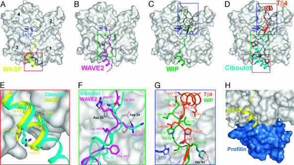
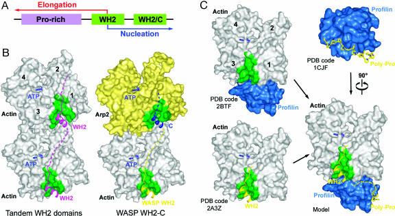
Wiskott-Aldrich syndrome protein (WASP)-homology domain 2 (WH2) is a small and widespread actin-binding motif. In the WASP family, WH2 plays a role in filament nucleation by Arp2/3 complex. Here we describe the crystal structures of complexes of actin with the WH2 domains of WASP, WASP-family verprolin homologous protein, and WASP-interacting protein. Despite low sequence identity, WH2 shares structural similarity with the N-terminal portion of the actin monomer-sequestering thymosin beta domain (Tbeta). We show that both domains inhibit nucleotide exchange by targeting the cleft between actin subdomains 1 and 3, a common binding site for many unrelated actin-binding proteins. Importantly, WH2 is significantly shorter than Tbeta but binds actin with approximately 10-fold higher affinity. WH2 lacks a C-terminal extension that in Tbeta4 becomes involved in monomer sequestration by interfering with intersubunit contacts in F-actin. Owing to their shorter length, WH2 domains connected in tandem by short linkers can coexist with intersubunit contacts in F-actin and are proposed to function in filament nucleation by lining up actin subunits along a filament strand. The WH2-central region of WASP-family proteins is proposed to function in an analogous way by forming a special class of tandem repeats whose function is to line up actin and Arp2 during Arp2/3 nucleation. The structures also suggest a mechanism for how profilin-binding Pro-rich sequences positioned N-terminal to WH2 could feed actin monomers directly to WH2, thereby playing a role in filament elongation.


Figures

Fig. 1.
Fig. 1.
Sequences and actin-binding affinities of WH2 and Tβ.(A) Domain structure of WASP (B, basic region; GBD, GTPase-binding domain; C, C region; A, acidic region). (B) Sequence alignment of Tβ and WH2. The sequence accession numbers are Q546P5 (Tβ4), O97428 (ciboulot), P18281 (actobindin), O43312 (MIM), Q53TA9 (WIP), P50552 (VASP), P42768 (WASP), O00401 (N-WASP), Q9Y6W5 (WAVE2), and Q9U4F1 (Spire). Conserved amino acids are colored according to their chemical characteristics (green, hydrophobic; blue, basic; red, acidic; yellow, small amino acid). The insert in the linker region of Tβ is shown in pink. Consensus profilin-binding sequences are shown in purple. Alignment of the CA region underlines the similarity between the C region and WH2. (C) Actin-binding affinities determined by ITC (see also Table 2). Highlighted in green are the WH2 domains whose structures were determined, whereas the amino acids that are seen in the structures are shown in bold in B.
Fig. 2.
Fig. 2.
WH2–actin structures. (AC) Structures of the WH2 domains of WASP, WAVE2, and WIP determined as ternary complexes with actin (gray) and DNase I (see Fig. 5). (D) Superimposition of the structures of ciboulot (9) and Tβ4 (10), which together represent Tβ–actin. (EG) Close-view comparisons of different parts of the WH2–actin and Tβ–actin structures shown in AD. (H) Partial overlap between the actin-binding sites of profilin (blue) and WH2.
Fig. 3.
Fig. 3.
Inhibition of nucleotide exchange on actin by WH2 and Tβ. ε-ATP–actin (1 μM) was mixed with 0, 0.5, 1, 2, 4, or 8 μM WASP430–458 (▪), WAVE433–464 (♦), WIP29–60 (▴), WIP29–46 (▵), MIM724–755 (×), Tβ42–44 (□), or Tβ42–33 (♦), and the fluorescence decay was monitored after addition of 1 mM ATP (see Materials and Methods).
Fig. 4.
Fig. 4.
Structural basis for the role of WH2 in filament nucleation and elongation. (A) In most proteins WH2 occurs in the form of tandem repeats C-terminal to Pro-rich sequences that support the binding of profilin–actin. The structures of WH2–actin suggest that this basic arrangement may allow WH2 to function in filament nucleation and elongation, two processes that are tightly connected but that can be conceptually separated. (B) Tandem repeats of short WH2 domains connected by short linkers can function to line up actin monomers along a filament strand, thereby playing a role in filament nucleation. Given the existing similarity between the C region of WASP/WAVE and WH2 (see Fig. 1B), the WH2 C region (C) may represent a specialized form of tandem repeat whose role is to add an actin subunit at the barbed end of Arp2 during Arp2/3 nucleation. (C) A superimposition of the structures of profilin–actin (19) and profilin–polyPro (36) with the structures of WH2–actin illustrates how profilin bound to the last consensus profilin-binding site (27) could deliver its actin directly to WH2, playing a role in filament elongation. A partial overlap between the actin-binding sites of profilin and WH2 may then help release profilin from the barbed end of the growing filament. Such a mechanism may constitute the basis for the so-called actoclampin model of actin polymerization (35).

References

    1. Pollard, T. D. & Borisy, G. G. (2003) Cell 112, 453–465. - PubMed
    1. Symons, M., Derry, J. M., Karlak, B., Jiang, S., Lemahieu, V., McCormick, F., Francke, U. & Abo, A. (1996) Cell 84, 723–734. - PubMed
    1. Bompard, G. & Caron, E. (2004) J. Cell Biol. 166, 957–962. - PMC - PubMed
    1. Machesky, L. M. & Insall, R. H. (1998) Curr. Biol. 8, 1347–1356. - PubMed
    1. Moreau, V., Frischknecht, F., Reckmann, I., Vincentelli, R., Rabut, G., Stewart, D. & Way, M. (2000) Nat. Cell. Biol. 2, 441–448. - PubMed

Publication types