. 2005 Nov 15;102(46):16584-9.
doi: 10.1073/pnas.0508306102. Epub 2005 Nov 7.

Resistance to inhibitors of cholinesterase 8A catalyzes release of Galphai-GTP and nuclear mitotic apparatus protein (NuMA) from NuMA/LGN/Galphai-GDP complexes

Affiliations

Resistance to inhibitors of cholinesterase 8A catalyzes release of Galphai-GTP and nuclear mitotic apparatus protein (NuMA) from NuMA/LGN/Galphai-GDP complexes

Gregory G Tall et al. Proc Natl Acad Sci U S A. .

Abstract

Resistance to inhibitors of cholinesterase (Ric) 8A is a guanine nucleotide exchange factor that activates certain G protein alpha-subunits. Genetic studies in Caenorhabditis elegans and Drosophila melanogaster have placed RIC-8 in a previously uncharacterized G protein signaling pathway that regulates centrosome movements during cell division. Components of this pathway include G protein subunits of the Galphai class, GPR or GoLoco domain-containing proteins, RGS (regulator of G protein signaling) proteins, and accessory factors. These proteins interact to regulate microtubule pulling forces during mitotic movement of chromosomes. It is unclear how the GTP-binding and hydrolysis cycle of Galphai functions in the context of this pathway. In mammals, the GoLoco domain-containing protein LGN (GPSM2), the LGN- and microtubule-binding nuclear mitotic apparatus protein (NuMA), and Galphai regulate a similar process. We find that mammalian Ric-8A dissociates Galphai-GDP/LGN/NuMA complexes catalytically, releasing activated Galphai-GTP in vitro. Ric-8A-stimulated activation of Galphai caused concomitant liberation of NuMA from LGN. We conclude that Ric-8A efficiently utilizes GoLoco/Galphai-GDP complexes as substrates in vitro and suggest that Ric-8A-stimulated release of Galphai-GTP and/or NuMA regulates the microtubule pulling forces on centrosomes during cell division.


Figures

Fig. 1.
Fig. 1.
Purification of the LGN/Gαi-1/NuMA (LGN BD) complex. (A) A soluble Sf9 cell lysate containing rat His-6-LGN was supplemented with purified myristoylated Gαi-1 from E. coli and rapidly adsorbed to Ni-NTA agarose. The resin was eluted with imidazole, and a portion of the eluate was resolved by SDS/PAGE; the gel was stained with Coomassie brilliant blue. (B) The eluate containing LGN/Gαi-1 was immediately supplemented with a 5-fold molar excess (to LGN) of myristoylated Gαi-1, loaded onto a Hi-trap Q column (GE Healthcare), and resolved with a linear NaCl gradient. Fractions of the eluate were resolved by SDS/PAGE. The fractions that were pooled are noted (Q pool). (C) The Q pool of LGN/Gαi-1 complex was incubated with a 4-fold molar excess of NuMA (LGN BD) and a molar equivalent of myristoylated-Gαi-1, ultracentrifuged, and gel-filtered over tandem Superdex 75/200 columns. Fractions of the gel filtration eluate were resolved by SDS/PAGE and stained. Fractions containing the LGN/Gαi-1/NuMA complex were pooled (Complex Pool) and used for subsequent assays.
Fig. 2.
Fig. 2.
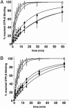
GTP[γS] binding to GoLoco protein/Gαi-1 complexes. (A) Full-length LGN/Gαi-1 (triangles), LGN/Gαi-1/NuMA (squares) complexes (50 nM), and free Gαi-1 (circles) (200 nM) were used to examine intrinsic (filled symbols) and 200 nM Ric-8A-stimulated (open symbols) rates of GTP[γS] binding. (B) LGNshort/Gαi-1 (triangles), AGS3short/Gαi-1 (squares) complexes (50 nM), and free Gαi-1 (circles) (200 nM) were used to examine intrinsic (filled symbols) and 200 nM Ric-8A-stimulated (open symbols) rates of GTP[γS] binding. Each reaction contained 10 mM MgCl2 and 10 μM[35S]GTP[γS] (10,000 cpm/pmol). Results are shown as the mean of triplicate experiments ± standard deviation.
Fig. 3.
Fig. 3.
Release of GDP from complexes containing myristoylated and unmodified Gαi-1 and LGNshort. Myristoylated Gαi-1 and unmodified Gαi-1 (100 nM) were bound to [α-32P]GDP and incubated with LGNshort (0, ▪; 5 nM, □;10 nM, •; 50 nM, ○; 100 nM, ▴; 200 nM, ▵; 500 nM, ▾ ;1 μM, ▿;5 μM, ♦) at 4°C for 5 min. Release of GDP from unmodified Gαi-1 was initiated at 30°C by adding 0 (A) or 200 nM (B) Ric-8A. Release of GDP from myristoylated Gαi-1 was initiated at 25°C by adding 0 (C) or 200 nM (D) Ric-8A. Duplicate aliquots of each reaction mixture were removed and quenched at the indicated times with AlF4 quench buffer. Results are shown as the mean of duplicate experiments (± standard deviation). The intrinsic (E) and Ric-8A-stimulated (F) rates of GDP release from myristoylated Gαi(•) and unmodified Gαi (○) are plotted logarithmically against LGNshort concentration by using origin 6.0 (Microcal Software).
Fig. 4.
Fig. 4.
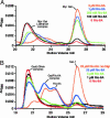
Ric-8A activates the LGNshort/myristoylated Gαi-1 complex catalytically. (A) Myristoylated Gαi-1/LGNshort complex (≈5 μM) was incubated with 50 μM GTP[γS], ≈1 mM free Mg+2, and 0 (red), 100 nM (black), 500 nM (green), 1 μM (blue), or 5 μM (orange) Ric-8A for 15 min at 25°C. (B) Unmodified Gαi-1/LGNshort complex (≈5 μM) was incubated with 50 μM GTP[γS], ≈1 mM free Mg+2, and 0 (red), 1 μM (black), 5 μM (green), or 25 μM (blue) Ric-8A for 15 min at 25°C or ≈10 mM free Mg+2 and 25 μM Ric-8A (orange) for 15 min at 30°C. Reaction mixtures were loaded onto tandem Superdex 75/200 columns and resolved. The UV absorbance traces of the column eluates are shown.
Fig. 5.
Fig. 5.
Ric-8A dissociates the LGN/Gαi-1/NuMA complex, activating myristoylated Gαi-1 and liberating NuMA. LGN/Gαi-1/NuMA complex (10 μM, ≈40 μM Gαi-1) was incubated for 10 (red) or 30 (blue) min at 30°C with 100 μM GDP (A), 100 μM GTP[γS] (B), 100 μM GDP and 1 μM Ric-8A (C), or 100 μM GTP[γS] and 1 μM Ric-8A (D). The reaction mixtures were loaded onto tandem Superdex 75/200 columns and resolved. Fractions from each experiment were analyzed by SDS/PAGE. Each silver-stained gel is shown below the UV absorbance of the gel filtration eluate. The positions of gel filtration mass standards and the fractions where NuMA dimer appeared are noted.

Similar articles

Cited by

References

    1. Manning, D. R. (2003) Sci. STKE 2003, pe35. - PubMed
    1. Goldstein, B. (2003) Curr. Biol. 13, R879–R880. - PubMed
    1. Hampoelz, B. & Knoblich, J. A. (2004) Cell 119, 453–456. - PubMed
    1. Siderovski, D. P. & Willard, F. S. (2005) Int. J. Biol. Sci. 1, 51–66. - PMC - PubMed
    1. Gotta, M. & Ahringer, J. (2001) Nat. Cell Biol. 3, 297–300. - PubMed

Publication types