. 2005 Nov 15;102(46):16777-82.
doi: 10.1073/pnas.0508303102. Epub 2005 Nov 7.

WNK3 kinase is a positive regulator of NKCC2 and NCC, renal cation-Cl- cotransporters required for normal blood pressure homeostasis

Affiliations

WNK3 kinase is a positive regulator of NKCC2 and NCC, renal cation-Cl- cotransporters required for normal blood pressure homeostasis

Jesse Rinehart et al. Proc Natl Acad Sci U S A. .

Abstract

WNK1 and WNK4 [WNK, with no lysine (K)] are serine-threonine kinases that function as molecular switches, eliciting coordinated effects on diverse ion transport pathways to maintain homeostasis during physiological perturbation. Gain-of-function mutations in either of these genes cause an inherited syndrome featuring hypertension and hyperkalemia due to increased renal NaCl reabsorption and decreased K(+) secretion. Here, we reveal unique biochemical and functional properties of WNK3, a related member of the WNK kinase family. Unlike WNK1 and WNK4, WNK3 is expressed throughout the nephron, predominantly at intercellular junctions. Because WNK4 is a potent inhibitor of members of the cation-cotransporter SLC12A family, we used coexpression studies in Xenopus oocytes to investigate the effect of WNK3 on NCC and NKCC2, related kidney-specific transporters that mediate apical NaCl reabsorption in the thick ascending limb and distal convoluted tubule, respectively. In contrast to WNK4's inhibitory activity, kinase-active WNK3 is a potent activator of both NKCC2 and NCC-mediated transport. Conversely, in its kinase-inactive state, WNK3 is a potent inhibitor of NKCC2 and NCC activity. WNK3 regulates the activity of these transporters by altering their expression at the plasma membrane. Wild-type WNK3 increases and kinase-inactive WNK3 decreases NKCC2 phosphorylation at Thr-184 and Thr-189, sites required for the vasopressin-mediated plasmalemmal translocation and activation of NKCC2 in vivo. The effects of WNK3 on these transporters and their coexpression in renal epithelia implicate WNK3 in NaCl, water, and blood pressure homeostasis, perhaps via signaling downstream of vasopressin.


Figures
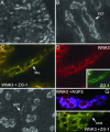
Fig. 1.
Fig. 1.
WNK3 is expressed at intercellular junctions along the nephron. Frozen mouse kidney sections were stained with anti-WNK3 antibody as described in Methods and visualized with immunofluorescence light microscopy. (A) WNK3 is present in all nephron segments. Low-power view of renal tubules in cross section stained with anti-WNK3 antibody reveals expression of WNK3 in all nephron segments. It is apparent that WNK3 predominantly localizes to intercellular junctions. (Original magnification, ×200.) (B) WNK3 expression in PCT. Tubule segments were stained with anti-WNK3 antibody and identified by costaining adjacent sections with anti-Megalin antibody, a PCT marker (not shown). (Original magnification, ×630.) (C-E) WNK3 (red) and ZO-1 (green) immunostaining in the TAL. Tubule segments were determined by costaining experiments with anti-WNK3 and anti-Tamm-Horsfall protein, a TAL marker (data not shown). WNK3 staining overlaps with ZO-1, a marker of tight junctions. (Original magnification, ×630.) (F) WNK3 expression in the DCT and connecting tubule (CNT), as determined by costaining experiments with anti-WNK3 and anti-Calbindin D-28K, a DCT/CNT marker (data not shown). (Original magnification, ×400.) (G) WNK3 expression in the cortical CD (CCD), determined by costaining with anti-WNK3 (red) and anti-Aquaporin-2 (AQP2), a CCD marker (blue). (Original magnification, ×630.) (H) WNK3 (red) and ZO-1 (green) immunostaining in the CCD. Tubule segments were determined by costaining experiments with anti-WNK3 and anti-AQP2. WNK3 staining overlaps with ZO-1, a marker of tight junctions. (Original magnification, ×700.)
Fig. 2.
Fig. 2.
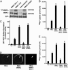
WNK3 kinase regulates the renal NCC and NKCC2 cotransporters. (A) WNK3 is an active kinase. Wild-type WNK3 (WT), kinase-dead WNK3 (kin-), and PHAII-like mutant WNK3 constructs were tagged with HA and expressed in COS-7 cells, purified by immunoprecipitation, and incubated with γ-32P ATP. The products were separated by SDS/PAGE, exposed to film, and also separately stained with anti-HA antibodies. Phosphorylated WNK3 (32P-WNK3) species were seen at 200 kDa (the size of WNK3-HA) in assays with WT and PHAII-like mutant WNK3 but were markedly reduced in assays with kinase-dead WNK3. Western blots with anti-HA and IgG demonstrate equivalent protein loading. (B) WNK3 regulates NCC. Xenopus oocytes were injected with cRNAs encoding NCC alone or in combination with wild-type WNK3, kinase-dead WNK3 (kin-), or PHAII-like WNK3. Metolazone-sensitive 22Na+ influx was measured as described in Methods. Results are expressed as mean ± SE of metolazone-sensitive 22Na+ influx. *, P < 0.0001 vs. NCC alone. WNK3 markedly increases metolazone-sensitive 22Na+ influx, kinase-dead WNK3 marked inhibits this activity, whereas PHAII-WNK3 behaves like wild-type WNK3. (C and D) WNK3 regulates NCC surface expression. (C) Oocytes were injected with cRNAs encoding EGFP-tagged NCC alone or in combination with wild-type or mutant WNK3. Surface expression of NCC was quantitated by confocal microscopy as described in Methods. Mean fluorescence seen in oocytes expressing GFP-NCC alone is expressed as 100%; other groups are expressed as a percentage of this value. Effects of WNK3 constructs on NCC surface expression closely parallel its effects on 22Na+ flux. *, P < 10-6 vs. NCC alone. (D) Examples of confocal microscopy of oocytes expressing GFP-tagged NCC alone or with wild-type or kinase-dead WNK3. (E) WNK3 regulates NKCC2. Oocytes were injected with cRNAs encoding NKCC2 alone or in combination with wild-type or mutant WNK3; bumetanide-sensitive 86Rb+ influx was measured as described in Methods. Mean ± SE of bumetanide-sensitive 86Rb+ influx is shown for each group in a representative experiment. As for NCC, WNK3 increases NKCC2 activity, kinase-dead WNK3 inhibits NKCC2, activity and PHAII-like WNK3 behaves like wild-type kinase.
Fig. 3.
Fig. 3.
WNK3 modulates the phosphorylation of NKCC2. Xenopus oocytes were injected with the indicated constructs and incubated at varying extracellular osmolarities as indicated. After incubation oocytes were lysed and Western blotting was performed by using the R5 (anti-phospho-NKCC2) or T9 (anti-NKCC2) antibodies as described in Methods. Phosphorylation of NKCC2 normally increases from negligible levels under hypotonic conditions (200 mM) to complete phosphorylation under hypertonic conditions (380 mM). In contrast, coexpression of NKCC2 with kinase-active WNK3 results in robust phosphorylation of NKCC2 at all osmolarities. Expression of kinase-dead WNK3 results in marked reduction of NKCC2 phosphorylation under hyperosmolar conditions.
Fig. 4.
Fig. 4.
Proposed role of WNK3 in the kidney. By regulating apical NaCl entry in the TAL and DCT of the nephron, WNK3 could modulate NaCl and water reabsorption and therefore blood pressure. Kinase-active WNK3 might increase NaCl reabsorption, whereas kinase-inactive WNK3 (WNK3 kin-) might inhibit NaCl reabsorption. These active/inactive states of WNK3 may be achieved dynamically by ligands (e.g., downstream of vasopressin) binding to or dissociating from the kinase.

References

    1. Lifton, R. P., Gharavi, A. G. & Geller, D. S. (2001) Cell 104, 545-556. - PubMed
    1. Simon, D. B., Nelson-Williams, C., Bia, M. J., Ellison, D., Karet, F. E., Molina, A. M., Vaara, I., Iwata, F., Cushner, H. M., Koolen, M., et al. (1996) Nat. Genet. 12, 24-30. - PubMed
    1. Simon, D. B., Karet, F. E., Hamdan, J. M., DiPietro, A., Sanjad, S. A. & Lifton R. P. (1996) Nat. Genet. 13, 183-188. - PubMed
    1. Seldin, D. W. & Giebisch, G. H. (2000) The Kidney: Physiology and Pathophysiology (Lippincott Williams & Wilkins, Philadelphia), 3rd Ed.
    1. Wilson, F. H., Disse-Nicodeme, S., Choate, K. A., Ishikawa, K., Nelson-Williams, C., Desitter, I., Gunel, M., Milford, D. V., Lipkin, G. W., Achard, J. M., et al. (2001) Science 293, 1107-1112. - PubMed

Publication types

Substances