. 2005 Nov;115(11):3185-92.
doi: 10.1172/JCI25444.

Synaptic activity becomes excitotoxic in neurons exposed to elevated levels of platelet-activating factor

Affiliations

Synaptic activity becomes excitotoxic in neurons exposed to elevated levels of platelet-activating factor

Matthew J Bellizzi et al. J Clin Invest. 2005 Nov.

Abstract

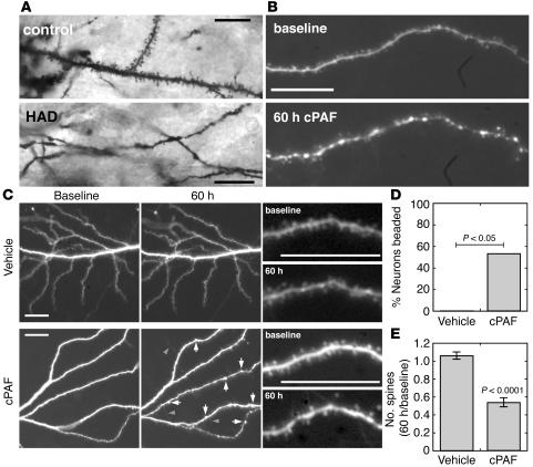
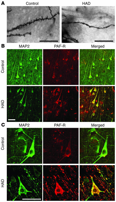
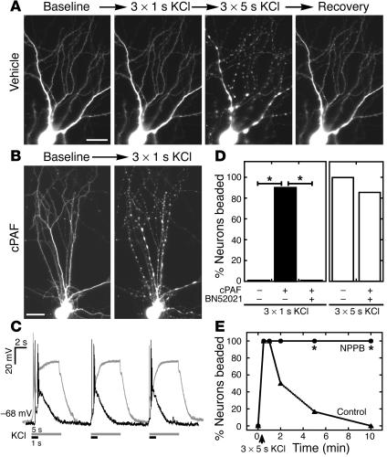
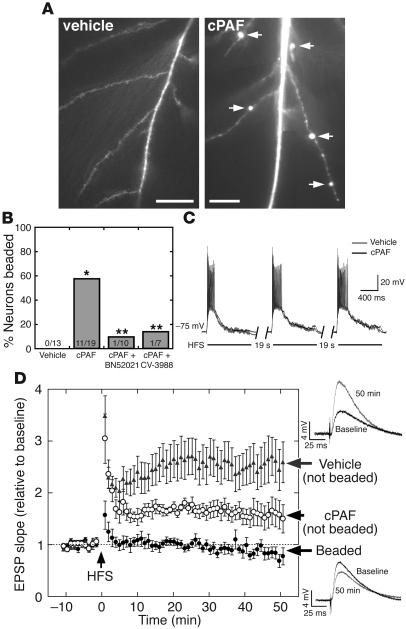
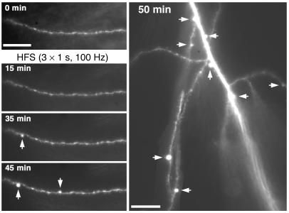
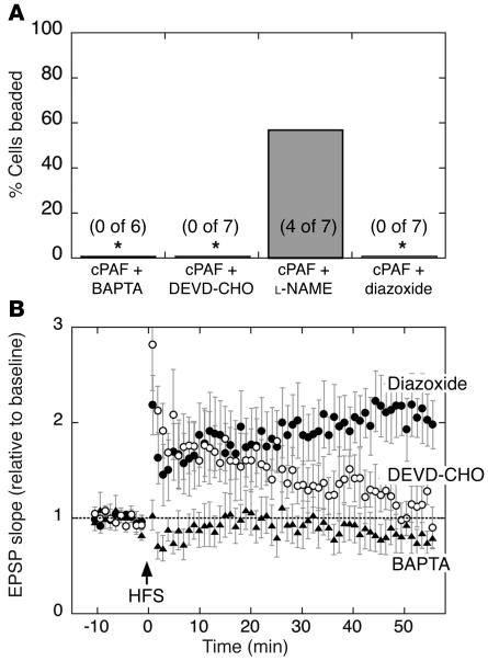
Neurologic impairment in HIV-1-associated dementia (HAD) and other neuroinflammatory diseases correlates with injury to dendrites and synapses, but how such injury occurs is not known. We hypothesized that neuroinflammation makes dendrites susceptible to excitotoxic injury following synaptic activity. We report that platelet-activating factor, an inflammatory phospholipid that mediates synaptic plasticity and neurotoxicity and is dramatically elevated in the brain during HAD, promotes dendrite injury following elevated synaptic activity and can replicate HIV-1-associated dendritic pathology. In hippocampal slices exposed to a stable platelet-activating factor analogue, tetanic stimulation that normally induces long-term synaptic potentiation instead promoted development of calcium- and caspase-dependent dendritic beading. Chemical preconditioning with diazoxide, a mitochondrial ATP-sensitive potassium channel agonist, prevented dendritic beading and restored long-term potentiation. In contrast to models invoking excessive glutamate release, these results suggest that physiologic synaptic activity may trigger excitotoxic dendritic injury during chronic neuroinflammation. Furthermore, preconditioning may represent a novel therapeutic strategy for preventing excitotoxic injury while preserving physiologic plasticity.


Figures

Figure 1
Figure 1
cPAF reproduces dendritic pathology of HAD. (A) Golgi-stained neurons in brain tissue from patients with HAD show focal swellings and fewer dendritic spines than those from HIV-1 seropositive controls without neurologic disease. (B) Dendrites in dissociated hippocampal cultures developed similar focal swellings and decreased numbers of spines after prolonged exposure to cPAF. (C) Lower-magnification images (left, middle columns) show dendritic beading (arrows) accompanied by sprouting of filopodia (arrowheads) with preservation of dendrite branches in cPAF-treated cultures and minimal change in dendrite morphology in vehicle-treated cultures. Higher-power images from the same cells (right column) show dendritic spine numbers maintained in a control dendrite and loss of spines in a cPAF-treated dendrite. (D) Of cPAF-treated neurons, 56% developed dendritic beading while none of the vehicle-treated cells did (n = 17, P < 0.05). (E) Numbers of dendritic spines decreased by 45% ± 5% with cPAF treatment and remained stable in control neurons (n = 10, P < 0.0001). Scale bars: 20 μm.
Figure 2
Figure 2
Dendritic injury and neuronal PAF-R expression in HAD. In cortical tissue from patients with HAD (A), dendritic beading and spine loss in Golgi-stained neurons is associated with (B) strong PAF-R immunohistochemical staining on dendrites and neuronal cell bodies identified by coimmunostaining for MAP2. HAD tissue shows fewer MAP2-positive dendritic branches compared with tissue from HIV-1 seropositive controls while PAF-R expression on the remaining dendrites and cell bodies is increased. (C) Higher-power field shows intense PAF-R expression on beaded dendrites in HAD compared with control dendrites. Scale bars: 20 μm.
Figure 3
Figure 3
cPAF increases vulnerability to dendritic swelling following synaptic activity. (A) In control cultures, synaptic activity due to 1-second depolarizing pulses of KCl elicited no change in dendrite morphology while 5-second pulses triggered beading throughout the dendritic arbor, which recovered within 10 minutes. (B) In cPAF-exposed cells, 1-second pulses caused rapid dendritic beading. (C) Membrane potential recordings show bursts of action potentials and stronger, more prolonged depolarization elicited by 5-second (white) versus 1-second (black) KCl stimulation. (D) cPAF lowered the threshold for activity-induced dendritic beading, leading to beading in 90% of neurons following 1-second KCl pulses that caused no beading in control neurons. PAF-R antagonist BN52021 blocked the increase in vulnerability. (E) Rapid recovery of dendritic beading is prevented by NPPB, an inhibitor of regulatory volume decrease in swollen neurons. *P < 0.001. Scale bars: 20 μm.
Figure 4
Figure 4
cPAF replaces LTP with dendritic beading in hippocampal slices. (A) Dendritic beading (arrows) in a cPAF-exposed CA1 pyramidal neuron 45 minutes after high-frequency Schaffer collateral stimulation (HFS) with no disruption of dendrite or spine morphology in following HFS in vehicle-treated cells. (B) HFS elicited dendritic beading in 11 of 19 cells from cPAF-treated slices and in 0 of 13 cells from vehicle-treated slices. *P < 0.001. PAF-R antagonists BN52021 and CV-3988 reduced dendritic beading to 1 of 10 and 1 of 7 cells, respectively. **P < 0.05 vs. cPAF. (C) The amplitude and duration of postsynaptic depolarization during HFS is unaffected by cPAF exposure. (D) Excitatory synaptic transmission is strongly potentiated following HFS in vehicle-treated slices (2.66 ± 0.44–fold relative to baseline at 40 to 50 minutes, n = 13, P < 0.001). In cPAF-treated slices, cells that did not develop dendritic beading underwent a smaller but significant potentiation (1.60 ± 0.26 relative to baseline, n = 8, P < 0.05) while EPSPs in cells whose dendrites did bead were not potentiated at all (0.84 ± 0.12 relative to baseline, n = 11, P < 0.01 vs. vehicle and P < 0.05 vs. cPAF-treated cells without dendritic beading). Representative EPSPs from vehicle-treated (upper right) and beaded cPAF-treated cells (lower right) are averages of 10 consecutive traces recorded at baseline and 50 minutes after HFS. Scale bars: 20 μm.
Figure 5
Figure 5
Activity-dependent dendritic beading is delayed, long lasting, and local. Dendritic beading in a cPAF-exposed hippocampal slice developed with a delay after HFS and progressed throughout the recording trial. In addition, focal swellings were restricted to discrete regions along the dendrite with no apparent disruption of dendrite and spine morphology in intervening areas. Scale bars: 20 μm.
Figure 6
Figure 6
Chemical preconditioning prevents calcium- and caspase-dependent beading and restores LTP. (A) Rates of dendritic beading and (B) EPSP potentiation following high-frequency Schaffer collateral stimulation in hippocampal slices exposed to cPAF. Postsynaptic calcium chelation by intracellularly applied BAPTA eliminated dendritic beading as well as synaptic potentiation (n = 6). Postsynaptic inhibition of caspase-3, -6, -7, -9, and -10 by intracellular Ac-DEVD-CHO (DEVD-CHO) (10 μM) prevented dendritic beading but failed to restore a lasting potentiation (n = 7) while nitric oxide synthase inhibitor L-NAME had no effect on rates of dendritic beading compared with cPAF alone (Figure 4). Pretreatment with the mitochondrial KATP agonist diazoxide prevented dendritic beading and restored LTP in cPAF-exposed slices (2.10 ± 0.27–fold potentiation at 40 to 50 minutes, n = 7). *P < 0.01 vs. cPAF alone (Figure 4).

References

    1. Masliah E, et al. Dendritic injury is a pathological substrate for human immunodeficiency virus-related cognitive disorders. HNRC Group. The HIV Neurobehavioral Research Center. Ann. Neurol. 1997;42:963–972. - PubMed
    1. Everall IP, Glass JD, McArthur J, Spargo E, Lantos P. Neuronal density in the superior frontal and temporal gyri does not correlate with the degree of human immunodeficiency virus-associated dementia. Acta Neuropathol. (Berl.). 1994;88:538–544. - PubMed
    1. Adle-Biassette H, et al. Neuronal apoptosis does not correlate with dementia in HIV infection but is related to microglial activation and axonal damage. Neuropathol. Appl. Neurobiol. 1999;25:123–133. - PubMed
    1. DeKosky ST, Scheff SW. Synapse loss in frontal cortex biopsies in Alzheimer’s disease: correlation with cognitive severity. Ann. Neurol. 1990;27:457–464. - PubMed
    1. Terry RD, et al. Physical basis of cognitive alterations in Alzheimer’s disease: synapse loss is the major correlate of cognitive impairment. Ann. Neurol. 1991;30:572–580. - PubMed

Publication types

MeSH terms