. 2006 Mar 1;394(Pt 2):399-407.
doi: 10.1042/BJ20051249.

Crystal structures of the editing domain of Escherichia coli leucyl-tRNA synthetase and its complexes with Met and Ile reveal a lock-and-key mechanism for amino acid discrimination

Affiliations

Crystal structures of the editing domain of Escherichia coli leucyl-tRNA synthetase and its complexes with Met and Ile reveal a lock-and-key mechanism for amino acid discrimination

Yunqing Liu et al. Biochem J. .

Abstract

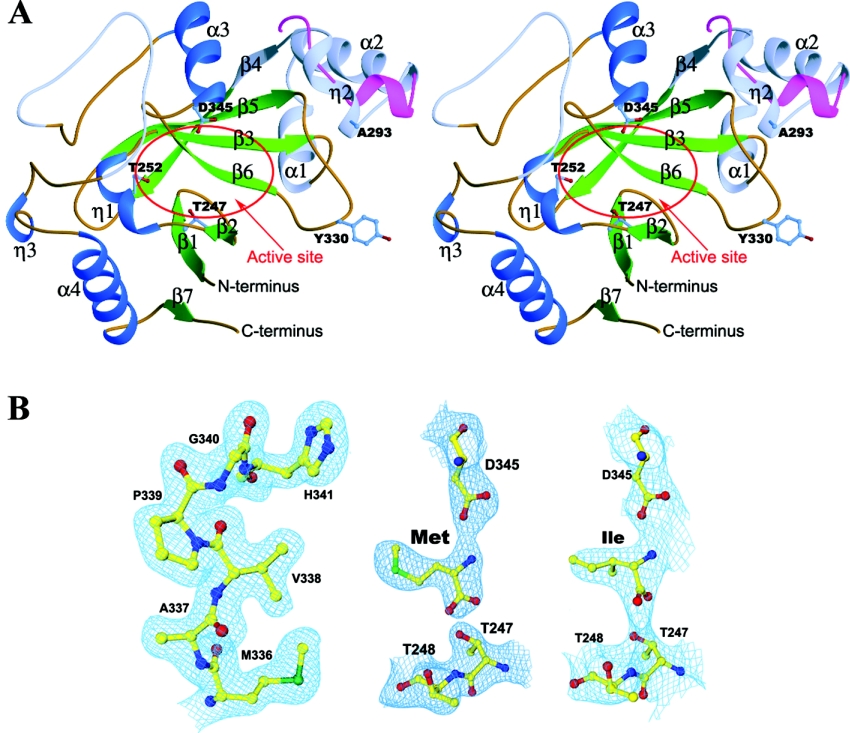
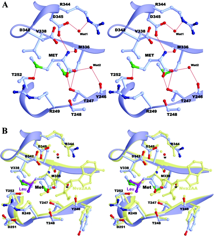
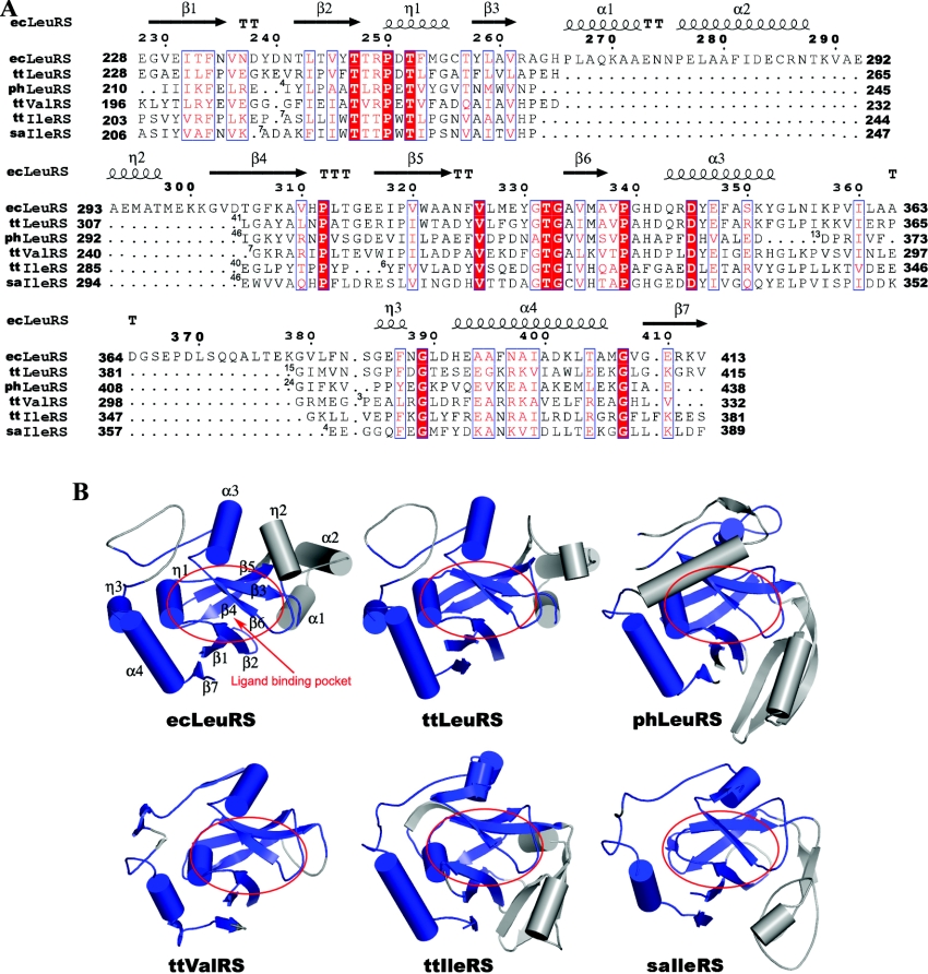
aaRSs (aminoacyl-tRNA synthetases) are responsible for the covalent linking of amino acids to their cognate tRNAs via the aminoacylation reaction and play a vital role in maintaining the fidelity of protein synthesis. LeuRS (leucyl-tRNA synthetase) can link not only the cognate leucine but also the nearly cognate residues Ile and Met to tRNA(Leu). The editing domain of LeuRS deacylates the mischarged Ile-tRNA(Leu) and Met-tRNA(Leu). We report here the crystal structures of ecLeuRS-ED (the editing domain of Escherichia coli LeuRS) in both the apo form and in complexes with Met and Ile at 2.0 A, 2.4 A, and 3.2 A resolution respectively. The editing active site consists of a number of conserved amino acids, which are involved in the precise recognition and binding of the noncognate amino acids. The substrate-binding pocket has a rigid structure which has an optimal stereochemical fit for Ile and Met, but has steric hindrance for leucine. Based on our structural results and previously available biochemical data, we propose that ecLeuRS-ED uses a lock-and-key mechanism to recognize and discriminate between the amino acids. Structural comparison also reveals that all subclass Ia aaRSs share a conserved structure core consisting of the editing domain and conserved residues at the editing active site, suggesting that these enzymes may use a common mechanism for the editing function.


Figures

Figure 1
Figure 1. Structure of ecLeuRS-ED
(A) Stereoview of the overall structure of ecLeuRS-ED and its comparison with ttLeuRS-ED. The α-helices, β-strands, and coils are coloured in blue, green and brown respectively. The non-conserved regions are coloured in light blue. The three strictly conserved residues at the editing active site (Thr247, Thr252, and Asp345) are shown with side chains. The major structural difference between ecLeuRS-ED and ttLeuRS-ED (in red) occurs in the Ala293-containing region. (B) Representative SIGMAA-weighted 2FoFc composite omit maps (1.0 σ contour level) at the editing active site. Left panel: apo form ecLeuRS-ED structure at 2.0 Å resolution; middle panel: the Met-bound complex at 2.4 Å resolution; and right panel: the Ile-bound complex at 3.2 Å resolution. The final coordinates of the structures are shown as ball-and-stick models.
Figure 2
Figure 2. Structure of the editing active site
(A) The Met-bound ecLeuRS-ED complex. The hydrogen-bonding interactions between Met and the surrounding residues are indicated with thin red lines. (B) Superposition of ecLeuRS-ED in complexes with Met (in light blue) and docked Leu (in purple) and ttLeuRS in complex with the post-transfer analogue Nva2AA (in light yellow).
Figure 3
Figure 3. Comparison of the ED of ecLeuRS with other subclass Ia aaRSs with known structures
(A) Structure-based sequence alignment of the ED between ecLeuRS and other subclass Ia aaRSs. ecLeuRS, ttLeuRS, phLeuRS, ttValRS, ttIleRS, and saIleRS refer to the editing domain of LeuRS in E. coli, T. thermophilus, and P. horikoshii, ValRS in T. thermophilus, and IleRS in T. thermophilus and S. aureus respectively. Invariant residues are highlighted in shaded red boxes and conserved residues in open red boxes. The secondary structure of ecLeuRS-ED is placed on top of the alignment. The numbers of residues omitted in the alignment are indicated by numbers at the beginning of each gap. The alignment was drawn with ESPript [32]. (B) Structural comparison of ecLeuRS-ED with the editing domain of other subclass Ia aaRSs. The conserved and non-conserved regions are coloured in blue and grey, respectively. The ligand binding pocket is indicated by red circles.
Figure 4
Figure 4. Docking model of the ecLeuRS-ED–tRNA complex
(A) The docking model of ttLeuRS in complex with tRNA. The subdomains of ttLeuRS are coloured as the RF domain and the small helical domain in green, ED in dark green, and LeuRS specific domain in red. The structure of ecLeuRS-ED (in purple) is superimposed onto the editing domain of ttLeuRS. tRNA is shown as light blue ribbon and the 3′ terminal CCA are shown with nucleotide bases. (B) Comparison of the 3′ terminal ACCA of the tRNA between the ValRS–tRNA complex (in blue) and the docking model of the ecLeuRS-ED–tRNA complex (in green). (C) Molecular surface of the Met-bound ecLeuRS-ED structure viewing from the entrance to the editing active site. Both the bound Met substrate and the docked 3′ terminal CCA of the tRNA are shown as ball-and-stick models.

Similar articles

Cited by

References

    1. Woese C., Olsen G. J., Ibba M., Soll D. Aminoacyl-tRNA synthetases, the genetic code, and the evolutionary process. Microbiol. Mol. Biol. Rev. 2000;64:202–236. - PMC - PubMed
    1. Freist W., Sternbach H., Cramer F. Isoleucyl-tRNA synthetase from baker's yeast and from Escherichia coli MRE 600. Discrimination of 20 amino acids in aminoacylation of tRNA(Ile)-C-C-A. Eur. J. Biochem. 1988;173:27–34. - PubMed
    1. Carter C. W., Jr Cognition, mechanism, and evolutionary relationships in aminoacyl-tRNA synthetases. Annu. Rev. Biochem. 1993;62:715–748. - PubMed
    1. Giege R., Sissler M., Florentz C. Universal rules and idiosyncratic features in tRNA identity. Nucleic Acids Res. 1998;26:5017–5035. - PMC - PubMed
    1. Nureki O., Vassylyev D. G., Tateno M., Shimada A., Nakama T., Fukai S., Konno M., Hendrickson T. L., Schimmel P., Yokoyama S. Enzyme structure with two catalytic sites for double-sieve selection of substrate. Science (Washington, D.C.) 1998;280:578–582. - PubMed

Publication types

LinkOut - more resources