

Copper chelation with tetrathiomolybdate suppresses adjuvant-induced arthritis and inflammation-associated cachexia in rats
- PMID: 16277669
- PMCID: PMC1297562
- DOI: 10.1186/ar1801
Copper chelation with tetrathiomolybdate suppresses adjuvant-induced arthritis and inflammation-associated cachexia in rats
Abstract
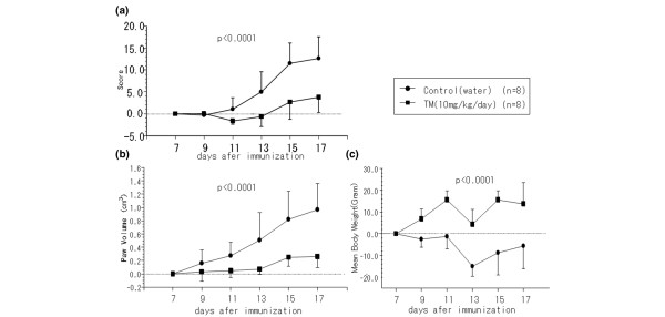
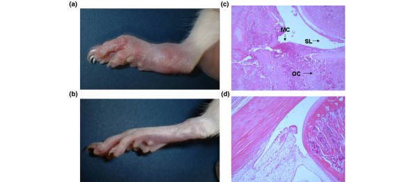
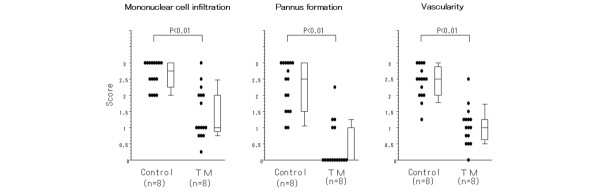
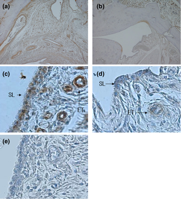
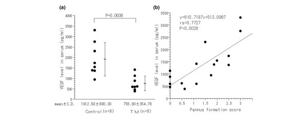
Tetrathiomolybdate (TM), a drug developed for Wilson's disease, produces an anti-angiogenic and anti-inflammatory effect by reducing systemic copper levels. TM therapy has proved effective in inhibiting the growth of tumors in animal tumor models and in cancer patients. We have hypothesized that TM may be used for the therapy of rheumatoid arthritis and have examined the efficacy of TM on adjuvant-induced arthritis in the rat, which is a model of acute inflammatory arthritis and inflammatory cachexia. TM delayed the onset of and suppressed the severity of clinical arthritis on both paw volume and the arthritis score. Histological examination demonstrated that TM significantly reduces the synovial hyperplasia and inflammatory cell invasion in joint tissues. Interestingly, TM can inhibit the expression of vascular endothelial growth factor in serum synovial tissues, especially in endothelial cells and macrophages. Moreover, the extent of pannus formation, which leads to bone destruction, is correlated with the content of vascular endothelial growth factor in the serum. There was no mortality in TM-treated rat abnormalities. TM also suppressed inflammatory cachexia. We suggest that copper deficiency induced by TM is a potent approach both to inhibit the progression of rheumatoid arthritis with minimal adverse effects and to improve the well-being of rheumatoid arthritis patients.
Figures
Similar articles
-
Protection against cartilage and bone destruction by systemic interleukin-4 treatment in established murine type II collagen-induced arthritis.Arthritis Res. 1999;1(1):81-91. doi: 10.1186/ar14. Epub 1999 Oct 26. Arthritis Res. 1999. PMID: 11056663 Free PMC article.
-
Tetrathiomolybdate is effective in a mouse model of arthritis.J Rheumatol. 2006 Dec;33(12):2501-6. Epub 2006 Oct 15. J Rheumatol. 2006. PMID: 17143984
-
Treatment with a neutralizing anti-murine interleukin-17 antibody after the onset of collagen-induced arthritis reduces joint inflammation, cartilage destruction, and bone erosion.Arthritis Rheum. 2004 Feb;50(2):650-9. doi: 10.1002/art.20001. Arthritis Rheum. 2004. PMID: 14872510
-
Cancer therapy with tetrathiomolybdate: antiangiogenesis by lowering body copper--a review.Integr Cancer Ther. 2002 Dec;1(4):327-37. doi: 10.1177/1534735402238185. Integr Cancer Ther. 2002. PMID: 14664727 Review.
-
The promise of copper lowering therapy with tetrathiomolybdate in the cure of cancer and in the treatment of inflammatory disease.J Trace Elem Med Biol. 2014 Oct;28(4):372-8. doi: 10.1016/j.jtemb.2014.07.015. Epub 2014 Aug 10. J Trace Elem Med Biol. 2014. PMID: 25194954 Review.
Cited by
-
Copper Promotes LPS-Induced Inflammation via the NF-кB Pathway in Bovine Macrophages.Biol Trace Elem Res. 2024 Dec;202(12):5479-5488. doi: 10.1007/s12011-024-04107-6. Epub 2024 Feb 20. Biol Trace Elem Res. 2024. PMID: 38376728
-
Hypoxia, cuproptosis, and osteoarthritis: Unraveling the molecular crosstalk.Redox Biol. 2025 Sep;85:103757. doi: 10.1016/j.redox.2025.103757. Epub 2025 Jul 8. Redox Biol. 2025. PMID: 40669206 Free PMC article.
-
Effect of allopurinol and vitamin e on rat model of rheumatoid arthritis.Int J Health Sci (Qassim). 2008 Jan;2(1):59-67. Int J Health Sci (Qassim). 2008. PMID: 21475473 Free PMC article.
-
Antiallergic and Antiarthritic Effects of Stem Bark Extract of Glyphaea brevis (Spreng) Monachino (Tiliaceae) in Murine Models.ISRN Pharmacol. 2013 Sep 17;2013:874263. doi: 10.1155/2013/874263. eCollection 2013. ISRN Pharmacol. 2013. PMID: 24167739 Free PMC article.
-
Tetrathiomolybdate Treatment Leads to the Suppression of Inflammatory Responses through the TRAF6/NFκB Pathway in LPS-Stimulated BV-2 Microglia.Front Aging Neurosci. 2018 Feb 27;10:9. doi: 10.3389/fnagi.2018.00009. eCollection 2018. Front Aging Neurosci. 2018. PMID: 29535623 Free PMC article.
References
-
- Firestein GS. Invasive fibroblast-like synoviocytes in rheumatoid arthritis: passive responders or transformed aggressors? Arthritis Rheum. 1996;39:1781–1790. - PubMed
Publication types
MeSH terms
Substances
Grants and funding
LinkOut - more resources
Full Text Sources
Other Literature Sources
