. 2005;7(6):R1189-99.
doi: 10.1186/ar1804. Epub 2005 Aug 17.

The contact-mediated response of peripheral-blood monocytes to preactivated T cells is suppressed by serum factors in rheumatoid arthritis

Affiliations

The contact-mediated response of peripheral-blood monocytes to preactivated T cells is suppressed by serum factors in rheumatoid arthritis

Manuela Rossol et al. Arthritis Res Ther. 2005.

Abstract

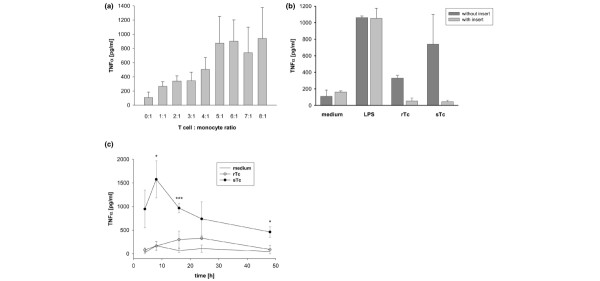
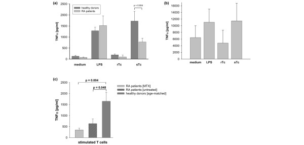
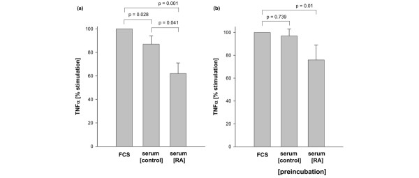
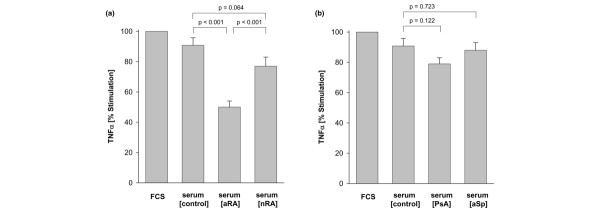
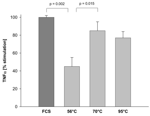
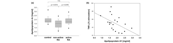
Stimulation of monocytes/macrophages after cell contact with preactivated T cells has been suggested to contribute to the excessive TNF-alpha production in rheumatoid arthritis (RA). In this study, T cell-contact-dependent TNF-alpha production by peripheral-blood monocytes in vitro was investigated and found to be significantly lower in treated and untreated patients with RA than in healthy controls. This suppression was not due to a general deficiency of monocytes to respond, because responses to lipopolysaccharide were comparable in patients and controls. In agreement with the pivotal role of TNF-alpha in RA, T cell-dependent induction of TNF-alpha in synovial macrophages was fivefold to tenfold higher than in peripheral-blood monocytes from either patients or controls. The decreased response of peripheral-blood monocytes from patients with RA was found to be mediated by inhibitory serum factors, because the addition of patient sera to monocytes from healthy controls suppressed TNF-alpha response in the co-culture assay. Preincubation of monocytes from healthy controls with RA serum was sufficient to suppress the subsequent TNF-alpha response in T cell co-cultures, indicating that inhibitory factors do indeed bind to monocyte surfaces, which might represent a regulatory counter-action of the immune system to the long-standing and consuming autoimmune process in RA. There are some indications that apolipoprotein A-1 might be part of this regulatory system.


Figures

Figure 1
Figure 1
T cell induced production of TNF-α by monocytes from healthy donors. (a) Fixed stimulated T cells induce tumour necrosis factor (TNF)-α production by monocytes in a dose-dependent manner. Peripheral-blood T cells (106/ml) were cultured for 48 hours in the presence of immobilized anti-CD3 (3.3 μg/ml) and soluble anti-CD28 (0.8 μg/ml) antibodies. Stimulated T cells were fixed and incubated for 24 hours with freshly isolated monocytes (1.5 × 10⁶/ml) at the indicated ratios. Values are means ± SEM from four different experiments. (b) Cell–cell contact is necessary for T cell-induced production of TNF-α in monocytes. Peripheral-blood T cells (106/ml) were cultured for 48 hours in the presence or absence of immobilized anti-CD3 (3.3 μg/ml) and soluble anti-CD28 (0.8 μg/ml) antibodies. Stimulated (sTc) and resting (rTc) T cells were fixed and incubated for 24 hours with freshly isolated monocytes (1.5 × 10⁶/ml) at a ratio of 7:1 in a transwell system as described in the Materials and methods section. T cells and monocytes were physically separated by the semi-permeable membrane (with insert) or had direct cell–cell contact (without insert). Lipopolysaccharide (LPS; 100 ng/ml) was used as a positive control. Values are means ± SEM for experiments with three different donors. (c) T cell-induced TNF-α production by monocytes is time-dependent. Peripheral-blood T cells (106/ml) were cultured for 48 hours in the presence or absence of immobilized anti-CD3 (3.3 μg/ml) and soluble anti-CD28 (0.8 μg/ml) antibodies. Stimulated (sTc) and resting (rTc) T cells were fixed and incubated for the indicated durations with freshly isolated monocytes (1.5 × 10⁶/ml) at a ratio of 7:1. Values are means ± SEM for experiments with three different donors. Levels of significance: * P < 0.05, *** P < 0.001.
Figure 2
Figure 2
Decreased T cell induced production of TNF-α in monocytes from RA patients. (a) T cell-induced tumour necrosis factor (TNF)-α secretion by monocytes from patients with rheumatoid arthritis (RA) is decreased in comparison with those from healthy controls. Peripheral-blood T cells (106/ml) were cultured for 48 hours in the presence or absence of immobilized anti-CD3 (3.3 μg/ml) and soluble anti-CD28 (0.8 μg/ml) antibodies. Stimulated (sTc) and resting (rTc) T cells were fixed and incubated with freshly isolated monocytes (1.5 × 10⁶/ml) at a ratio of 7:1. After 24 hours, the concentration of TNF-α was measured in the supernatant. Lipopolysaccharide (LPS; 100 ng/dl) was used as a positive control. Data are means ± SEM of values from 20 patients with RA and 20 age-matched controls. (b) Macrophages separated from the synovial membrane of patients with RA produce large amounts of TNF-α after contact with preactivated T cells. Stimulated (sTc) and resting (rTc) T cells were fixed and incubated with freshly isolated synovial macrophages from patients with RA (1.5 × 10⁶/ml) at a ratio of 7:1. After 24 hours, the concentration of TNF-α was measured in the supernatant. LPS (100 ng/ml) was used as a positive control. Data are means ± SEM of values from six independent experiments. Level of significance: not significant. (c) Decrease in T cell-induced TNF-α secretion by monocytes from patients with RA is independent of previous and current treatments. The graph depicts the T cell-induced TNF-α production by monocytes of patients with RA either receiving methotrexate (MTX; n = 5) or not receiving immunosuppressive treatment (untreated; n = 6). For comparison, results for six age-matched controls are given (P < 0.05, significant difference compared with untreated patients).
Figure 3
Figure 3
Sera from RA patients inhibit T cell-induced TNF-α production by monocytes from healthy donors. (a) Peripheral-blood T cells (106/ml) were cultured for 48 hours in the presence of immobilized anti-CD3 (3.3 μg/ml) and soluble anti-CD28 (0.8 μg/ml) antibodies. The cells were fixed and incubated for 24 hours with freshly isolated monocytes (1.5 × 10⁶/ml) at a ratio of 7:1. The co-incubation assay was performed in the presence of 10% FCS, 10% serum from patients (serum [RA]) or from healthy donors (serum [control]). All data are expressed as percentages of TNF-α produced in the 10% FCS containing co-culture system (100%). Data are means ± SEM for 10 independent experiments; levels of significance are as indicated. (b) Monocytes from healthy donors were preincubated with 50% control sera (nine donors) or RA sera (six sera from patients with RA, which were previously found to inhibit T cell-dependent monocyte activation) and then washed thoroughly three times. Co-culture experiments were performed as described in the text in medium supplemented with 10% FCS. All data are expressed as percentages of TNF-α produced in the co-culture system containing 10% FCS. Data are means ± SEM for four independent experiments; levels of significance are as indicated.
Figure 4
Figure 4
Inhibition of T cell-induced TNF-α production by monocytes is specific for active RA. (a) Preactivated peripheral-blood T cells (106/ml; see Materials and methods) were fixed and incubated for 24 hours with freshly isolated peripheral-blood monocytes (1.5 × 10⁶/ml) at a ratio of 7:1. The co-incubation assay was performed in the presence of 10% FCS, 10% serum from healthy donors (serum [control], n = 10), from patients with active RA (serum [aRA], n = 20) or from patients with non-active RA (serum [nRA], n = 20). All data are expressed as percentages of TNF-α produced in the co-culture system containing 10% FCS. Data are means ± SEM; levels of significance are as indicated. (b) Co-incubation assays (see (a)) were performed in the presence of 10% FCS, 10% serum from healthy donors (serum [control], n = 10)), serum from patients with psoriatic arthritis (serum [PsA], n = 9) or from patients with ankylosing spondylitis (serum [aSp], n = 9). All data are expressed as percentages of TNF-α produced in the co-culture system containing 10% FCS. Data are means ± SEM; levels of significance are as indicated.
Figure 5
Figure 5
Inhibitory activity of rheumatoid arthritis (RA) sera is not heat stable. Six sera from patients with RA, which previously were found to strongly inhibit T cell-dependent monocyte activation were incubated at 56°C for 30 min, at 70°C for 10 min or at 95°C for 2 min. Co-culture experiments were performed in the presence of 10% FCS or 10% RA sera. All data are expressed as percentages of tumour necrosis factor-α produced in the co-culture system containing 10% FCS. Data are means ± SEM from six independent experiments. P = 0.002 (significant difference from the 100% values).
Figure 6
Figure 6
ApoA-1 concentrations are decreased in non-active RA but correlate with the inhibitory serum activity in active RA. (a) Box plot depicting ApoA-1 concentrations in sera from healthy controls (n = 20), in patients with active RA (n = 20) and in patients with non-active disease (n = 20). Levels of significance are given; n.s., not significant. (b) Scatter plot depicting the correlation between ApoA-1 concentrations and inhibitory serum activity in sera from patients with active RA. Each data point represents the ApoA-1 concentration in relation to the inhibitory activity. Tumour necrosis factor (TNF)-α production in the co-culture system containing 10% FCS is set to 100%. The regression line illustrates the negative correlation between the two parameters (correlation coefficient R = -0.527; P = 0.016).

References

    1. Burger D. Cell contact-mediated signaling of monocytes by stimulated T cells: a major pathway for cytokine induction. Eur Cytokine Netw. 2000;11:346–353. - PubMed
    1. Isler P, Vey E, Zhang JH, Dayer JM. Cell surface glycoproteins expressed on activated human T cells induce production of interleukin-1 beta by monocytic cells: a possible role of CD69. Eur Cytokine Netw. 1993;4:15–23. - PubMed
    1. Manie S, Kubar J, Limouse M, Ferrua B, Ticchioni M, Breittmayer JP, Peyron JF, Schaffar L, Rossi B. CD3-stimulated Jurkat T cells mediate IL-1 beta production in monocytic THP-1 cells. Role of LFA-1 molecule and participation of CD69 T cell antigen. Eur Cytokine Netw. 1993;4:7–13. - PubMed
    1. Lecoanet-Henchoz S, Gauchat JF, Aubry JP, Graber P, Life P, Paul-Eugene N, Ferrua B, Corbi AL, Dugas B, Plater-Zyberk C, et al. CD23 regulates monocyte activation through a novel interaction with the adhesion molecules CD11b-CD18 and CD11c-CD18. Immunity. 1995;3:119–125. doi: 10.1016/1074-7613(95)90164-7. - DOI - PubMed
    1. Armant M, Rubio M, Delespesse G, Sarfati M. Soluble CD23 directly activates monocytes to contribute to the antigen-independent stimulation of resting T cells. J Immunol. 1995;155:4868–4875. - PubMed

Publication types

MeSH terms

Substances