. 2005;7(6):R1338-47.
doi: 10.1186/ar1833. Epub 2005 Sep 30.

Reduced transforming growth factor-beta signaling in cartilage of old mice: role in impaired repair capacity

Affiliations

Reduced transforming growth factor-beta signaling in cartilage of old mice: role in impaired repair capacity

E N Blaney Davidson et al. Arthritis Res Ther. 2005.

Abstract

Osteoarthritis (OA) is a common joint disease, mainly effecting the elderly population. The cause of OA seems to be an imbalance in catabolic and anabolic factors that develops with age. IL-1 is a catabolic factor known to induce cartilage damage, and transforming growth factor (TGF)-beta is an anabolic factor that can counteract many IL-1-induced effects. In old mice, we observed reduced responsiveness to TGF-beta-induced IL-1 counteraction. We investigated whether expression of TGF-beta and its signaling molecules altered with age. To mimic the TGF-beta deprived conditions in aged mice, we assessed the functional consequence of TGF-beta blocking. We isolated knee joints of mice aged 5 months or 2 years, half of which were exposed to IL-1 by intra-articular injection 24 h prior to knee joint isolation. Immunohistochemistry was performed, staining for TGF-beta1, -2 or -3, TGF-betaRI or -RII, Smad2, -3, -4, -6 and -7 and Smad-2P. The percentage of cells staining positive was determined in tibial cartilage. To mimic the lack of TGF-beta signaling in old mice, young mice were injected with IL-1 and after 2 days Ad-LAP (TGF-beta inhibitor) or a control virus were injected. Proteoglycan (PG) synthesis (35S-sulfate incorporation) and PG content of the cartilage were determined. Our experiments revealed that TGF-beta2 and -3 expression decreased with age, as did the TGF-beta receptors. Although the number of cells positive for the Smad proteins was not altered, the number of cells expressing Smad2P strongly dropped in old mice. IL-1 did not alter the expression patterns. We mimicked the lack of TGF-beta signaling in old mice by TGF-beta inhibition with LAP. This resulted in a reduced level of PG synthesis and aggravation of PG depletion. The limited response of old mice to TGF-beta induced-IL-1 counteraction is not due to a diminished level of intracellular signaling molecules or an upregulation of intracellular inhibitors, but is likely due to an intrinsic absence of sufficient TGF-beta receptor expression. Blocking TGF-beta distorted the natural repair response after IL-1 injection. In conclusion, TGF-beta appears to play an important role in repair of cartilage and a lack of TGF-beta responsiveness in old mice might be at the root of OA development.


Figures

Figure 1
Figure 1
Number of cells in medial and lateral tibial cartilage of 5 month and 2 year old C57Bl/6 mice. Paraffin sections of knee joints of young (5 months old) and old (2 years old) mice were stained with hematoxylin and eosin after which a blinded observer used a computerized imaging system to count the number of chondrocytes in the tibial cartilage. Old mice have a lower number of cells in their cartilage than young mice. The reduction in cell number is more pronounced on the medial side of the joint. Error bars display the standard error. For statistical analysis, a Student's t-test was used. * = p < 0.05; ** = p < 0.005; *** = p < 0.0005.
Figure 2
Figure 2
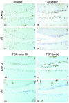
Staining of various transforming growth factor (TGF)-beta signaling molecules in cartilage. Paraffin sections of knee joints of young and old mice were stained with antibodies against (a,c) Smad2, (b,d) Smad-2P, (e,g) TGF-beta receptor II (TGF-betaRII) and (f,h) TGF-beta2. The medial tibia of the young mice clearly show a high number of cells staining positive for (b) Smad-2P, (e) TGF-betaRII and (f) TGF-beta2, whereas the (d,g,h) old mice had only a very low number of cells staining positive for these factors. (a,c) Smad2 staining remained unchanged with age. F, femur; T, tibia.
Figure 3
Figure 3
Percentage of cells expressing transforming growth factor (TGF)-beta in medial and lateral tibial cartilage. Paraffin sections of knee joints of young (5 months old) and old (2 years old) mice were stained immunohistochemically with antibodies against TGF-beta1, TGF-beta2 or TGF-beta3. Subsequently, the number of cells staining positive in the cartilage were scored with a computerized imaging system and corrected for the total number of cells. (a) In medial cartilage, TGF-beta2 and TGF-beta3 expression were significantly reduced with age. (b) In lateral cartilage, TGF-beta2 was significantly reduced. Error bars display the standard error. For statistical analysis, a Student's t-test was used. * = p < 0.05.
Figure 4
Figure 4
Percentage of cells expressing transforming growth factor (TGF)-beta receptors (TGF-betaRs). Paraffin sections of knee joints of young (5 months old) and old mice (2 years old) were stained immunohistochemically with antibodies against TGF-betaRI or TGF-betaRII. Subsequently, the number of cells staining positive in the cartilage were scored with a computerized imaging system and corrected for the total number of cells. The expression of both receptors was reduced with age in both (a) medial and (b) lateral tibial cartilage, but the reduced TGF-betaRII was significant only in lateral tibial cartilage. Error bars display the standard error. For statistical analysis, a Student's t-test was used. * = p < 0.05; ** = p < 0.005; *** = p < 0.0005.
Figure 5
Figure 5
Percentage of cells expressing Smad in medial and lateral tibial cartilage. Frozen sections of knee joints of young (5 months old) and old (2 years old) mice were stained immunohistochemically with antibodies against Smad2, Smad3, Smad4, Smad6 or Smad7. Subsequently, the number of cells staining positive were scored with a computerized imaging system and corrected for the total number of cells. (a) In medial tibial cartilage, expression of Smad3, Smad4 and Smad7 increased with age. (b) In lateral tibial cartilage Smad3 and Smad6 expression increased with age. Error bars display the standard error. For statistical analysis, a Student's t-test was used. * = p < 0.05; *** = p < 0.0005.
Figure 6
Figure 6
Percentage of cells expressing Smad-2P in medial and lateral tibial cartilage. Paraffin sections of knee joints of young (5 months old) and old (2 years old) mice were stained immunohistochemically with antibodies against Smad-2P. Subsequently, the number of cells staining positive were scored with a computerized imaging system and corrected for the total number of cells. The Smad-2P expression was reduced with age in both medial and lateral tibial cartilage. Error bars display the standard error. For statistical analysis, a Student's t-test was used. *** = p < 0.0005.
Figure 7
Figure 7
Effect of IL-1 on expression of TGF-beta signaling proteins in cartilage. Knee joints of (a) young (5 months old) and (b) old (2 years old) mice were injected with IL-1 24 h prior to isolation of the knee joints. Paraffin sections of knee joints were stained immunohistochemically for TGF-beta1, TGF-beta2, TGF-beta3, TGF-beta receptor I (TGF-betaRI), TGF-betaRII, Smad2, Smad3, Smad4, Smad6, Smad7 and Smad-2P. Subsequently, the number of cells staining positive were scored with a computerized imaging system and corrected for the total number of cells. After IL-1 injection, Smad2 expression increased only in the medial tibial cartilage and TGF-beta1 and TGF-beta2 expression increased only in the lateral tibial cartilage. Error bars display the standard error. For statistical analysis, a Student's t-test was used. * = p < 0.05.
Figure 8
Figure 8
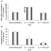
Effect of transforming growth factor (TGF)-beta deprivation on intrinsic cartilage repair capacity. Murine knee joints of young mice were injected with IL-1. After two days an adenovirus expressing the TGF-beta inhibitor latency associated peptide (LAP) was injected intra-articularly. Four days after the initial injections with IL-1, patellae were isolated for 35S-sulfate incorporation and whole knee joints were isolated for histology. (a) 35S-sulfate incorporation into isolated patellar cartilage after treatment with IL-1 and Ad-LAP. IL-1 treatment induces an initial decrease in 35S-sulfate incorporation, but by day 4 the 35S-sulfate incorporation increased above normal levels, indicating an overshoot in proteoglycan synthesis. By blocking endogenous TGF-beta with LAP, this overshoot is completely abolished. (b) Proteoglycan content of patellar cartilage after treatment with IL-1 and Ad-LAP. IL-1 injection results in depletion of proteoglycans in cartilage. Blocking endogenous TGF-beta with LAP results in an aggravation of this depletion beyond IL-1 induced damage alone.

Comment in

References

    1. Moos V, Fickert S, Muller B, Weber U, Sieper J. Immunohistological analysis of cytokine expression in human osteoarthritic and healthy cartilage. J Rheumatol. 1999;26:870–879. - PubMed
    1. Towle CA, Hung HH, Bonassar LJ, Treadwell BV, Mangham DC. Detection of interleukin-1 in the cartilage of patients with osteoarthritis: a possible autocrine/paracrine role in pathogenesis. Osteoarthritis Cartilage. 1997;5:293–300. - PubMed
    1. Webb GR, Westacott CI, Elson CJ. Osteoarthritic synovial fluid and synovium supernatants up-regulate tumor necrosis factor receptors on human articular chondrocytes. Osteoarthritis Cartilage. 1998;6:167–176. doi: 10.1053/joca.1998.0109. - DOI - PubMed
    1. van Beuningen HM, Arntz OJ, van den Berg WB. In vivo effects of interleukin-1 on articular cartilage. Prolongation of proteoglycan metabolic disturbances in old mice. Arthritis Rheum. 1991;34:606–615. - PubMed
    1. Ganu V, Goldberg R, Peppard J, Rediske J, Melton R, Hu SI, Wang W, Dunander C, Heingard D. Inhibition of interleukin-1alpha-induced cartilage oligomeric matrix protein degradation in bovine articular cartilage by matrix metalloproteinase inhibitors: potential role for matrix metalloproteinases in the generation of cartilage oligomeric matrix protein fragments in arthritic synovial fluid. Arthritis Rheum. 1998;41:2143–2151. doi: 10.1002/1529-0131(199812)41:12<2143::AID-ART9>3.0.CO;2-P. - DOI - PubMed

Publication types

MeSH terms