

Kinetic, stability, and structural changes in high-resolution crystal structures of HIV-1 protease with drug-resistant mutations L24I, I50V, and G73S
- PMID: 16277992
- PMCID: PMC1403828
- DOI: 10.1016/j.jmb.2005.09.095
Kinetic, stability, and structural changes in high-resolution crystal structures of HIV-1 protease with drug-resistant mutations L24I, I50V, and G73S
Abstract
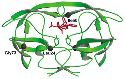
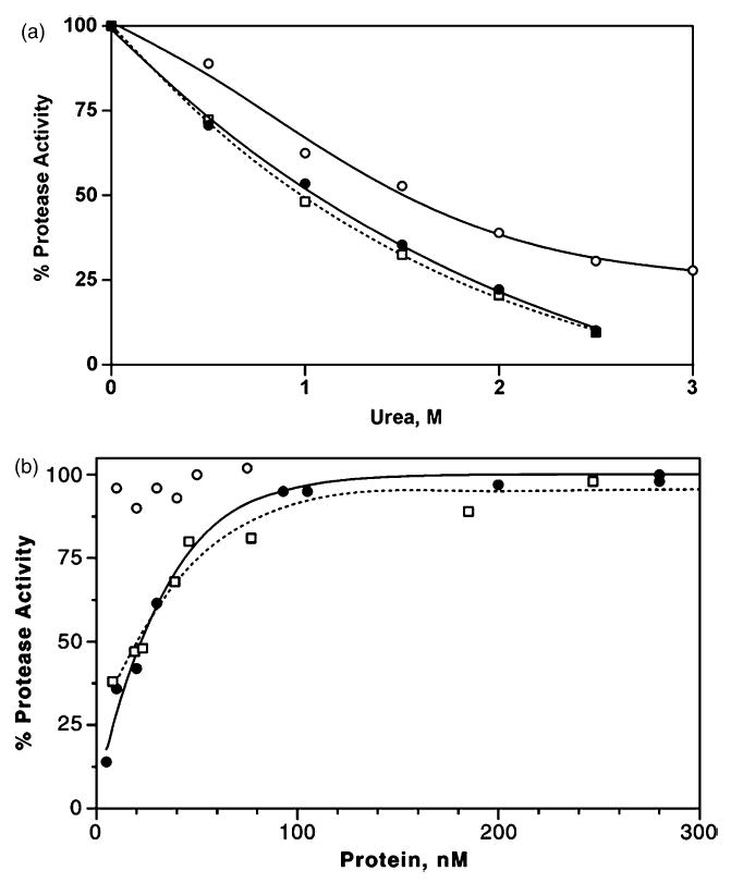
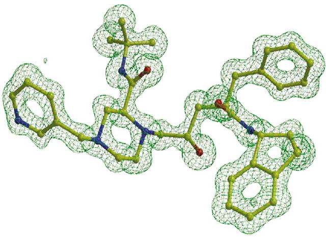
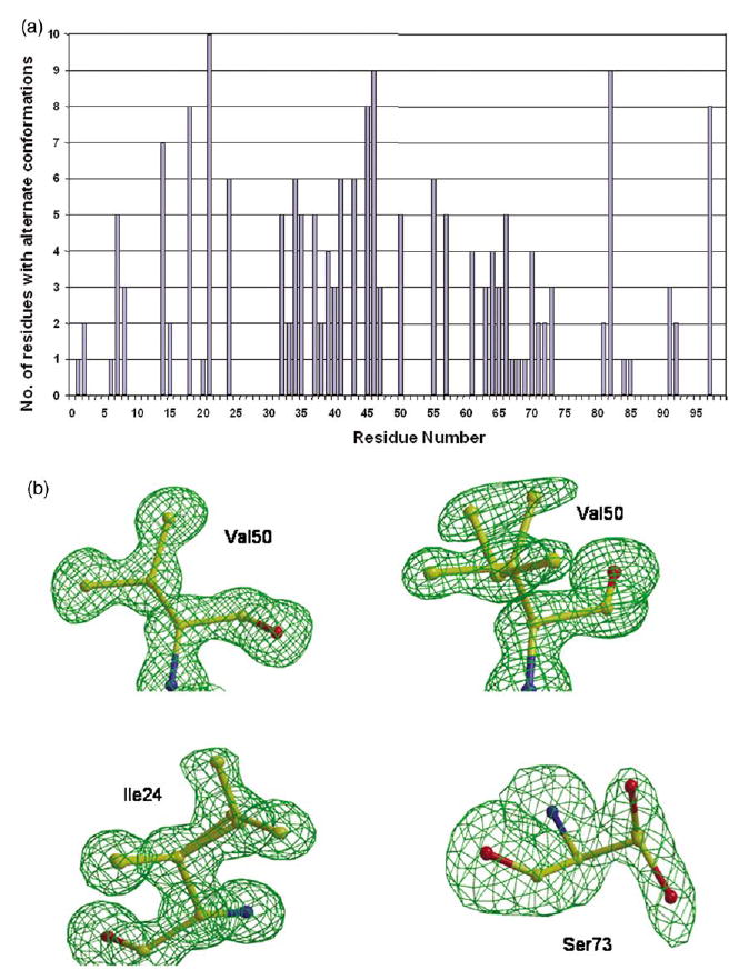
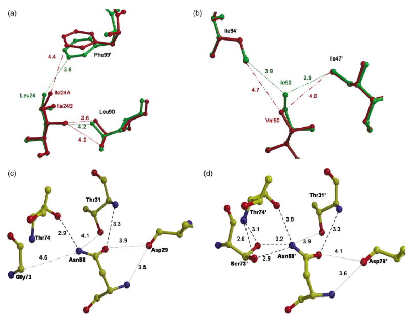
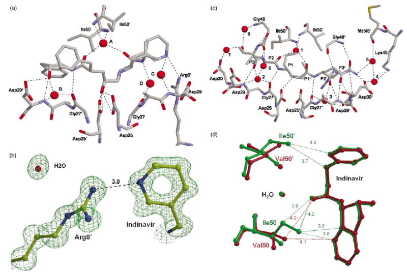
The crystal structures, dimer stabilities, and kinetics have been analyzed for wild-type human immunodeficiency virus type 1 (HIV-1) protease (PR) and resistant mutants PR(L24I), PR(I50V), and PR(G73S) to gain insight into the molecular basis of drug resistance. The mutations lie in different structural regions. Mutation I50V alters a residue in the flexible flap that interacts with the inhibitor, L24I alters a residue adjacent to the catalytic Asp25, and G73S lies at the protein surface far from the inhibitor-binding site. PR(L24I) and PR(I50V), showed a 4% and 18% lower k(cat)/K(m), respectively, relative to PR. The relative k(cat)/K(m) of PR(G73S) varied from 14% to 400% when assayed using different substrates. Inhibition constants (K(i)) of the antiviral drug indinavir for the reaction catalyzed by the mutant enzymes were about threefold and 50-fold higher for PR(L24I) and PR(I50V), respectively, relative to PR and PR(G73S). The dimer dissociation constant (K(d)) was estimated to be approximately 20 nM for both PR(L24I) and PR(I50V), and below 5 nM for PR(G73S) and PR. Crystal structures of the mutants PR(L24I), PR(I50V) and PR(G73S) were determined in complexes with indinavir, or the p2/NC substrate analog at resolutions of 1.10-1.50 Angstrom. Each mutant revealed distinct structural changes relative to PR. The mutated residues in PR(L24I) and PR(I50V) had reduced intersubunit contacts, consistent with the increased K(d) for dimer dissociation. Relative to PR, PR(I50V) had fewer interactions of Val50 with inhibitors, in agreement with the dramatically increased K(i). The distal mutation G73S introduced new hydrogen bond interactions that can transmit changes to the substrate-binding site and alter catalytic activity. Therefore, the structural alterations observed for drug-resistant mutations were in agreement with kinetic and stability changes.
Figures
Similar articles
-
Molecular basis for substrate recognition and drug resistance from 1.1 to 1.6 angstroms resolution crystal structures of HIV-1 protease mutants with substrate analogs.FEBS J. 2005 Oct;272(20):5265-77. doi: 10.1111/j.1742-4658.2005.04923.x. FEBS J. 2005. PMID: 16218957 Free PMC article.
-
Effect of flap mutations on structure of HIV-1 protease and inhibition by saquinavir and darunavir.J Mol Biol. 2008 Aug 1;381(1):102-15. doi: 10.1016/j.jmb.2008.05.062. Epub 2008 Jul 1. J Mol Biol. 2008. PMID: 18597780 Free PMC article.
-
Amprenavir complexes with HIV-1 protease and its drug-resistant mutants altering hydrophobic clusters.FEBS J. 2010 Sep;277(18):3699-714. doi: 10.1111/j.1742-4658.2010.07771.x. Epub 2010 Aug 2. FEBS J. 2010. PMID: 20695887 Free PMC article.
-
Effectiveness of nonpeptide clinical inhibitor TMC-114 on HIV-1 protease with highly drug resistant mutations D30N, I50V, and L90M.J Med Chem. 2006 Feb 23;49(4):1379-87. doi: 10.1021/jm050943c. J Med Chem. 2006. PMID: 16480273 Free PMC article.
-
A contribution to the drug resistance mechanism of darunavir, amprenavir, indinavir, and saquinavir complexes with HIV-1 protease due to flap mutation I50V: a systematic MM-PBSA and thermodynamic integration study.J Chem Inf Model. 2013 Aug 26;53(8):2141-53. doi: 10.1021/ci4002102. Epub 2013 Jul 24. J Chem Inf Model. 2013. PMID: 23834142
Cited by
-
Conserved hydrogen bonds and water molecules in MDR HIV-1 protease substrate complexes.Biochem Biophys Res Commun. 2013 Jan 18;430(3):1022-7. doi: 10.1016/j.bbrc.2012.12.045. Epub 2012 Dec 19. Biochem Biophys Res Commun. 2013. PMID: 23261453 Free PMC article.
-
Unique Flap Conformation in an HIV-1 Protease with High-Level Darunavir Resistance.Front Microbiol. 2016 Feb 3;7:61. doi: 10.3389/fmicb.2016.00061. eCollection 2016. Front Microbiol. 2016. PMID: 26870021 Free PMC article.
-
Inhibition of XMRV and HIV-1 proteases by pepstatin A and acetyl-pepstatin.FEBS J. 2012 Sep;279(17):3276-86. doi: 10.1111/j.1742-4658.2012.08714.x. Epub 2012 Aug 17. FEBS J. 2012. PMID: 22804908 Free PMC article.
-
Accurate Prediction of Drug Activity by Computational Methods: Importance of Thermal Capacity.Molecules. 2025 Jun 12;30(12):2563. doi: 10.3390/molecules30122563. Molecules. 2025. PMID: 40572530 Free PMC article.
-
HIV-1 protease function and structure studies with the simplicial neighborhood analysis of protein packing method.Proteins. 2008 Nov 15;73(3):742-53. doi: 10.1002/prot.22094. Proteins. 2008. PMID: 18498108 Free PMC article.
References
-
- Hertogs K, Bloor S, Kemp SD, Van den Eynde C, Alcorn TM, Pauwels R, et al. Phenotypic and genotypic analysis of clinical HIV-1 isolates reveals extensive protease inhibitor cross-resistance: a survey of over 6000 samples. AIDS. 2000;14:1203–1210. - PubMed
-
- Louis JM, Weber IT, Tozser J, Clore GM, Gronenborn AM. HIV-1 protease: maturation, enzyme specificity, and drug resistance. Advan Pharmacol. 2000;49:111–146. - PubMed
-
- Ridky TW, Kikonyogo A, Leis J, Gulnik S, Copeland T, Erickson J, et al. Drug-resistant HIV-1 proteases identify enzyme residues important for substrate selection and catalytic rate. Biochemistry. 1998;37:13835–13845. - PubMed
Publication types
MeSH terms
Substances
Associated data
- Actions
- Actions
- Actions
- Actions
- Actions
Grants and funding
LinkOut - more resources
Full Text Sources
Chemical Information
Research Materials
Miscellaneous
