. 2005 Dec;17(12):3362-77.
doi: 10.1105/tpc.105.036889. Epub 2005 Nov 8.

Interactions between the cell cycle and embryonic patterning in Arabidopsis uncovered by a mutation in DNA polymerase epsilon

Affiliations
Free PMC article

Interactions between the cell cycle and embryonic patterning in Arabidopsis uncovered by a mutation in DNA polymerase epsilon

Pablo D Jenik et al. Plant Cell. 2005 Dec.
Free PMC article

Abstract

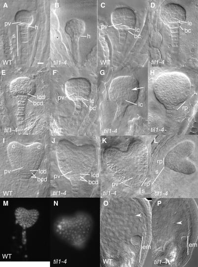
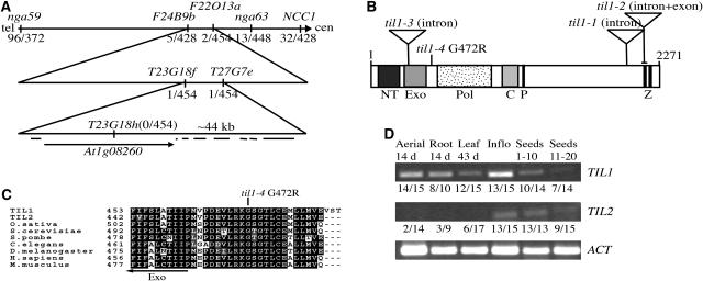
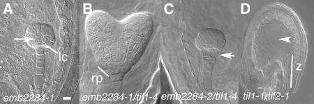
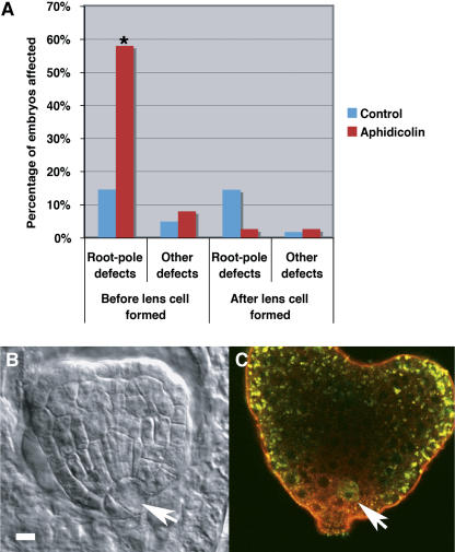
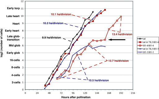
Pattern formation and morphogenesis require coordination of cell division rates and orientations with developmental signals that specify cell fate. A viable mutation in the TILTED1 locus, which encodes the catalytic subunit of DNA polymerase epsilon of Arabidopsis thaliana, causes a lengthening of the cell cycle by approximately 35% throughout embryo development and alters cell type patterning of the hypophyseal lineage in the root, leading to a displacement of the root pole from its normal position on top of the suspensor. Treatment of preglobular and early globular stages, but not later stage, embryos with the DNA polymerase inhibitor aphidicolin leads to a similar phenotype. The results uncover an interaction between the cell cycle and the processes that determine cell fate during plant embryogenesis.


Figures

Figure 1.
Figure 1.
Phenotypes of Wild-Type and Homozygous til1-4 Embryos. Note that in all cases, wild-type embryos are compared with til1-4/til1-4 embryos of similar embryonic stage. The til1-4/til1-4 embryos develop more slowly and are therefore always chronologically older than their wild-type counterparts. h, hypophysis; s, suspensor; lc, lens cell; lcd, lens cell descendant; bc, basal cell; bcd, basal cell descendants; pv, provascular cells; c, cotyledon; rp, root pole; em, embryo. Bars = 10 μm in (A) to (K), 15 μm in (L) to (N), and 20 μm in (O) and (P). (A) Wild-type early globular embryo. (B) til1-4/til1-4 early globular embryo. (C) Wild-type mid globular embryo. (D) and (E) til1-4/til1-4 mid globular embryos. In (D), the lens cell is asymmetric. In (E), both the lens cell and basal cell have divided longitudinally. (F) Wild-type late globular embryo. (G) and (H) til1-4/til1-4 late globular embryos. In (G), the lens cell is asymmetric. Cells in the right side of the embryo (arrow) show abnormal divisions. (I) Wild-type early heart stage embryo. The hypophyseal cell lineage is symmetric. (J) to (L) til1-4/til1-4 early heart stage embryos. In all three cases, the hypophyseal cell lineage is abnormal. The resulting tilt of the shoot–root axis relative to the suspensor axis varies from slight (J) to moderate (K) to extreme (L). (M) and (N) Wild-type (M) and til1-4/til1-4 (N) DAPI-stained early heart stage embryos. (O) and (P) Wild-type (O) and til1-4/til1-4 (P) free nuclear endosperm at 96 h after pollination.
Figure 2.
Figure 2.
Expression of Cell-Specific Markers in Wild-Type and Homozygous til1-4 Embryos. The root pole is shown at higher magnification in the insets in (C) to (F), (I) to (L), and (O) to (Q). h, hypophysis; lc, lens cell; lcd, lens cell descendants; bc, basal cell; bcd, basal cell descendants; ce, cortex-endodermis initials; e, endodermis. Bars = 10 μm in (A) to (D), (K) to (L), and (W) to (Z), 15 μm in (E), (F), (I), (J), and (O) to (Q), and 7.7 μm in (G), (H), (M), (N), and (R) to (V). (A) and (B) PSTM:GUS expression in wild-type (A) and til1-4/til1-4 (B) heart stage embryos. (C) to (F) PIN4:GUS expression in wild-type ([C] and [E]) and til1-4/til1-4 ([D] and [F]) embryos. (C), transition; (D), late globular; (E) and (F), late heart stages. In older mutant embryos (F), PIN4:GUS expression is asymmetrically offset relative to the suspensor. (G) to (L) DR5rev:GFP expression in wild-type ([G], early globular; [I], heart; [K], torpedo stages) and til1-4/til1-4 embryos ([H], early globular; [J], heart; [L], torpedo stages). In the sections in (J) and (L), a single lens cell descendant can be observed. (M) to (Q) QC25 expression in wild-type ([M], early heart; [O], torpedo stages) and til1-4/til1-4 mutant embryos ([N], early heart; [P] and [Q] torpedo stages). This expression highlights abnormalities in the lens cell lineage in mutants; the embryo in (N) has an abnormally shaped lens cell, while the embryo in (Q) has fewer lens cell descendants than the wild type. (R) to (Z) PSCR:GFP expression in wild-type ([R], early globular stage; [T], mid globular; [W], heart; [Y], torpedo stages) and til1-4/til1-4 embryos ([S], early globular; [U], late globular; [V], mid globular stages). Later in development, the normally U-shaped pattern of PSCR:GFP expression is often asymmetric ([X], heart; [Z], torpedo stages), reflecting the lateral displacement of the root pole.
Figure 3.
Figure 3.
TIL1 Encodes the Catalytic Subunit of DNA Pol ɛ. (A) Map position of the til1-4 mutation on chromosome I. The fractions shown indicate the number of recombinant chromosomes over the total number of chromosomes scored for each marker. (B) Schematic of the TIL1 protein, showing the organization of its domains (NT, N-terminal; Exo, 3′-5′ exonuclease; Pol, 5′-3′ polymerase; C, central; P, proliferating cell nuclear antigen interaction; Z, zinc-finger). The position of the T-DNA insertions (til1-1 to til1-3) and the point mutation (til1-4) are shown. (C) Alignment of the region surrounding the amino acid residue altered by the til1-4 mutation. (D) Expression of TIL1 and TIL2 as determined by RT-PCR. The numbers under each lane indicate the fraction of times a band was detected. Actin was used as a control. Inflo, inflorescence; seeds 1-10, seeds from the first 10 siliques (to late heart stage); seeds 11-20, seeds from the next 10 siliques (torpedo stage to fully developed embryo).
Figure 4.
Figure 4.
Phenotypes of Embryos Carrying til1 Insertion Alleles (emb2284) and of Double Mutants for til1-1 and til2-1. (A) emb2284-1/emb2284-1 embryo arrested at early globular stage. Arrow shows abnormal division patterns. It also has an asymmetric lens cell. (B) emb2284-1/til1-4 heart stage embryo with a laterally displaced root pole. (C) emb2284-2/til1-4 early globular embryo with a longitudinally split hypophysis (arrow). (D) Seed with a til1-1/til1-1;til2-1/til2-1 embryo arrested as a zygote and a single endosperm nucleus (arrowhead). lc, lens-shaped cell; rp, root pole; z, zygote. Bars = 10 μm in (A) to (D).
Figure 5.
Figure 5.
Treatment of Embryos with Aphidicolin. (A) Graph of the percentage of embryos affected by treatment with aphidicolin. Embryos at two-cell to early globular embryo stages at the beginning of treatment were grouped in the “before lens cell formed” (control, n = 144; aphidicolin, n = 113), while embryos at later stages (to late globular stage) were grouped in the “after lens cell formed class” (control, n = 55; aphidicolin, n = 39). The observed morphological abnormalities were separated into root pole defects (til1-4-like) and other defects (other parts of the embryo). Asterisk indicates a significant difference with the control group (P < 0.01, Fisher's exact test). (B) Transition stage embryo after aphidicolin treatement with an abnormally divided hypophysis (arrow). (C) Early heart embryo after aphidicolin treatment showing an abnormally positioned PSCR:GFP-expressing cell (arrow). The punctate green fluorescence is an artifact. Bar = 10 μm for (B) and (C).
Figure 6.
Figure 6.
Cell Number in Wild-Type and til Mutant Embryos during Embryogenesis. The three wild-type samples have similar cell doubling times. til1-1/til1-1 embryos arrest at mid globular stage. til1-4/til1-4 embryos develop slower than the wild type and pause at the late globular stage before continuing to develop (arrow). The data plotted are from Supplemental Table 1 online.

Similar articles

Cited by

References

    1. Aida, M., Beis, D., Heidstra, H., Willemsen, V., Blilou, I., Galinha, C., Nussaume, L., Noh, Y.-S., Amasino, R.M., and Scheres, B. (2004). The PLETHORA genes mediate patterning of the Arabidopsis root stem cell niche. Cell 119 109–120. - PubMed
    1. Alonso, J.M.A., et al. (2003). Genome-wide insertional mutagenesis of Arabidopsis thaliana. Science 301 653–657. - PubMed
    1. Ambros, V. (1999). Cell cycle-dependent sequencing of cell fate decisions in Caenorhabditis elegans vulva precursor cells. Development 126 1947–1956. - PubMed
    1. Barrôco, R.M., Van Poucke, K., Bergervoet, J.H.W., De Veylder, L., Groot, S.P.C., Inzé, D., and Engler, G. (2005). The role of the cell cycle machinery in resumption of postembryonic development. Plant Physiol. 137 127–140. - PMC - PubMed
    1. Berleth, T., and Jürgens, G. (1993). The role of the monopteros gene in organizing the basal body region of the Arabidopsis embryo. Development 118 575–587.

Publication types

Associated data