. 2005 Nov;4(11):1775-84.
doi: 10.1128/EC.4.11.1775-1784.2005.

Involvement of a short interspersed element in epigenetic transcriptional silencing of the amoebapore gene in Entamoeba histolytica

Affiliations

Involvement of a short interspersed element in epigenetic transcriptional silencing of the amoebapore gene in Entamoeba histolytica

Michael Anbar et al. Eukaryot Cell. 2005 Nov.

Abstract

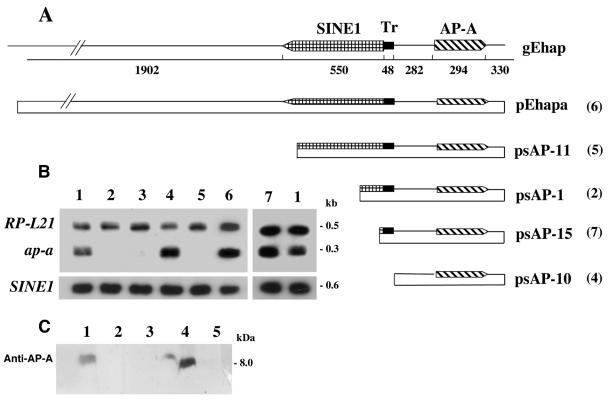
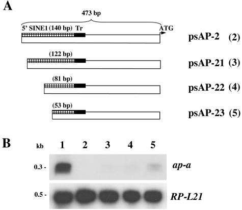
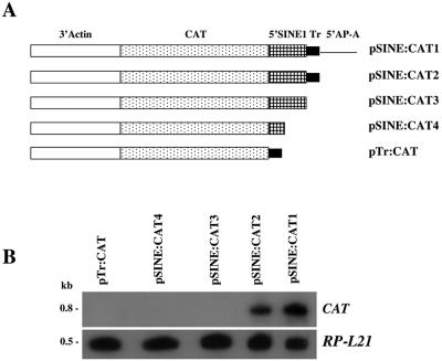
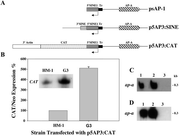
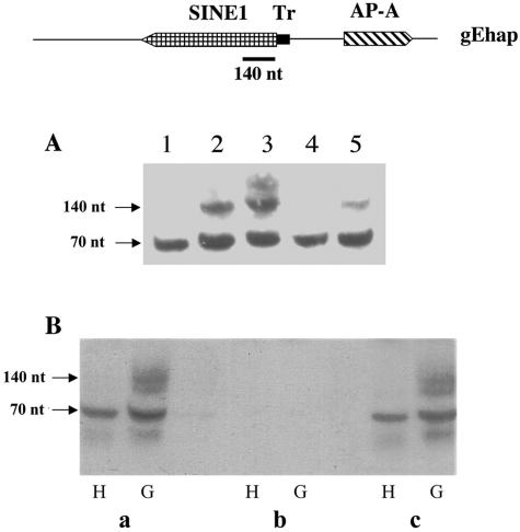
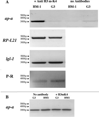
Transcriptional silencing of an amoebapore (ap-a) gene occurred in Entamoeba histolytica following the transfection of plasmids containing a DNA segment (473 bp) homologous to the 5' upstream region of the gene (R. Bracha, Y. Nuchamowitz, and D. Mirelman, Eukaryot. Cell 2:295-305, 2003). This segment contains the promoter region of the ap-a gene, a T-rich stretch, followed by a truncated SINE1 (short interspersed element 1) that is transcribed from the antisense strand. Transfection of plasmids containing truncated SINE1 sequences which lack their 3' regulatory elements upstream of the ap-a gene was essential for the downstream silencing of the ap-a gene while transfection with plasmids containing the entire SINE1 sequence or without the T-rich stretch promoted the overexpression of the ap-a gene. Both the T-rich stretch and sequences of the 5' SINE1 were essential for the transcription of SINE1. RNA extracts from gene-silenced cultures showed small amounts of short (approximately 140-nucleotide), single-stranded molecules with homology to SINE1 but no short interfering RNA. Chromatin immunoprecipitation analysis with an antibody against methylated K4 of histone H3 showed a demethylation of K4 at the domain of the ap-a gene, indicating transcriptional inactivation. These results suggest the involvement of SINE1 in triggering the gene silencing and the role of histone modification in its epigenetic maintenance.


Figures

FIG. 1.
FIG. 1.
(A) gEhap (top): schematic diagram of the amoebapore and SINE1 genomic region. Arrowed boxes represent transcription orientation. The T-rich (Tr) region is represented by a solid bar. Plasmids: diagrams of the different fragments cloned into the pEhActNeo shuttle vector (length corresponds to the scheme above). pEhapa is derived from gEhap (accession no. X-70851). Numbers in parentheses represent the corresponding transfected trophozoites. (B) Northern blot analysis of amoebic RNA extracts. Lane 1, (both ends) parent strain HM1:IMSS; lane 2, transfected psAP-1; lane 3, plasmid-less G3; lane 4, psAP-10; lane 5, psAP-11; lane 6, pEhapa; lane 7, psAP-15. Blots were probed with radiolabeled DNA of the ap-a coding region, ribosomal protein gene EhRP-L21, and SINE1 probe. (C) Western blot of SDS-polyacrylamide gel reacted with anti-AP-A antibodies (samples 6 and 7 not shown).
FIG. 2.
FIG. 2.
(A) Schematic map of various plasmid constructs containing different lengths of the 5′ flanking region of the ap-a gene. The length of the truncated 5′ SINE is given in parentheses. (B) Northern blots hybridized with radiolabeled probes of ap-a and ribosomal protein L21 ORFs. Lane 1, parent strain HM-1:IMSS; lane 2, psAP-2; lane 3, psAP-21; lane 4, psAP-22; lane 5, psAP-23.
FIG. 3.
FIG. 3.
(A) Schematic map of the various plasmid constructs containing the CAT reporter gene and 3′ regulatory element of actin fused to different upstream regions of SINE1. (B) Northern blot analysis. RNA was extracted from HM1:IMSS transfectants with the different plasmids, probed with the radiolabeled CAT gene, and subsequently probed with a radiolabeled ribosomal protein gene, EhRP-L21.
FIG. 4.
FIG. 4.
(A) Schematic map of the various chimeric plasmids used for transfection of parent strain HM-1:IMSS and ap-a-silenced G3 trophozoites. (B) Expression levels of CAT transcripts in trophozoites of strain HM-1:IMSS and silenced G3 which were transfected with p5AP3:CAT plasmid. Quantitative analysis of CAT RNA levels, which were normalized for plasmid copy number. The densitometric analysis was normalized by defining CAT/Neo ratios of expression in transfected strain HM-1:IMSS as 100%. (C) Northern blot analysis of amoebic RNA extracts. Lane 1, HM-1:IMSS; lane 2, p5AP3:CAT transfected into HM-1:IMSS; lane 3, p5AP3:CAT in G3. (D) Northern blot analysis of amoebic RNA extracts. Lane 1, HM-1:IMSS; lane 2, p5AP3:SINE transfected into HM-1:IMSS; lane 3, psAP-1 transfected into HM-1:IMSS. Blots in panels C and D were hybridized with radiolabeled probes of ap-a.
FIG. 5.
FIG. 5.
(A) Small RNA analysis on a 17% acrylamide gel. Samples are from the indicated trophozoite extracts: lane 1, HM-1:IMSS; lane 2, psAP-1; lane 3, G3; lane 4, psAP-10; lane 5, psAP-11. Blots were hybridized with a radiolabeled, top-strand-oriented probe derived from the 5′ 140 bp of SINE1 (top diagram). (B) The small RNA extracts from parent strain HM-1:IMSS (H) and silenced trophozoites G3 (G) were treated with RNA-modifying enzymes according to manufacturer's instructions before being loaded on the gel, and blots were hybridized as in panel A. (a) Intact samples; (b) RNase I treatment; (c) RNase III treatment.
FIG. 6.
FIG. 6.
(A) PCR amplification of genes from DNA fragments following ChIP with antibody against methylated K4 of histone H3 from trophozoites of strain HM-1:IMSS and ap-a gene-silenced G3. Precipitates without antibodies served as negative controls. Genes that were amplified by PCR before and after immunoprecipitation were amoebapore A (ap-a), ribosomal protein L21 (RP-L21), light subunit of Gal lectin (lgl-1), and a proline-rich gene (P-R) which neighbors another SINE (42). (B) DNA samples prior to immunoprecipitation were used as PCR templates and served as input controls. Primers used for PCR amplification of the different genes are shown in Table 1. For details on ChIP procedures see Materials and Methods.

References

    1. Abed, M., and S. Ankri. 2005. Molecular characterization of Entamoeba histolytica RNase III and AGO2, two RNA interference hallmark proteins. Exp. Parasitol. 110:265-269. - PubMed
    1. Ainbinder, E., M. Revach, O. Wolstein, S. Moshonov, N. Diamant, and R. Dikstein. 2002. Mechanism of rapid transcriptional induction of tumor necrosis factor alpha-responsive genes by NF-κB. Mol. Cell. Biol. 22:6354-6362. - PMC - PubMed
    1. Alon, R. N., R. Bracha, and D. Mirelman. 1997. Inhibition of expression of the lysine-rich 30 kDa surface antigen of Entamoeba dispar by the transcription of its antisense RNA. Mol. Biochem. Parasitol. 90:193-201. - PubMed
    1. Bakrea, A. A., K. Rawal, R. Ramaswamy, A. Bhattacharya, and S. Bhattacharya. 2005. The LINEs and SINEs of Entamoeba histolytica: comparative analysis and genomic distribution. Exp. Parasitol. 110:207-213. - PubMed
    1. Bannister, A. J., R. Schneider, and T. Kouzarides. 2002. Histone methylation: dynamic or static? Cell 109:801-806. - PubMed

Publication types