. 2005 Nov;4(11):1863-71.
doi: 10.1128/EC.4.11.1863-1871.2005.

Independent metalloregulation of Ace1 and Mac1 in Saccharomyces cerevisiae

Affiliations

Independent metalloregulation of Ace1 and Mac1 in Saccharomyces cerevisiae

Greg Keller et al. Eukaryot Cell. 2005 Nov.

Abstract

Ace1 and Mac1 undergo reciprocal copper metalloregulation in yeast cells. Mac1 is functional as a transcriptional activator in copper-deficient cells, whereas Ace1 is a transcriptional activator in copper-replete cells. Cells undergoing a transition from copper-deficient to copper-sufficient conditions through a switch in the growth medium show a rapid inactivation of Mac1 and a corresponding rise in Ace1 activation. Cells analyzed after the transition show a massive accumulation of cellular copper. Under these copper shock conditions we show, using two epitope-tagged variants of Mac1, that copper-mediated inhibition of Mac function is independent of induced protein turnover. The transcription activity of Mac1 is rapidly inhibited in the copper-replete cells, whereas chromatin immunoprecipitation studies showed only partial copper-induced loss of DNA binding. Thus, the initial event in copper inhibition of Mac1 function is likely copper inhibition of the transactivation activity. Copper inhibition of Mac1 in transition experiments is largely unaffected in cells overexpressing copper-binding proteins within the nucleus. Likewise, high expression of a copper-binding, non-DNA-binding Mac1 mutant is without effect on the copper activation of Ace1. Thus, metalloregulation of Ace1 and Mac1 occurs independently.


Figures
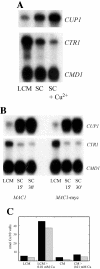
FIG. 1.
FIG. 1.
Expression of CUP1 and CTR1 as a function of the cellular copper status. Panel A. Steady-state cells were cultured overnight in SC containing the BIO101 low-copper nitrogen base. The culture was split into three aliquots and diluted into medium containing either LCM, SC, or SC plus 0.1 mM CuSO4. Cells were harvested and S1 nuclease analysis was carried out on purified RNA. The expression of CUP1 and CTR1 was assessed relative to CMD1 (encoding calmodulin) as the loading control. Panel B. Cells were cultured overnight in SC glucose containing the BIO101 low-copper nitrogen base. These cells were used to inoculate a culture in LCM (+BCS) for 5.5 h. Cells were then transitioned to SC medium and were harvested at either 15 or 30 min following the transition to SC medium. RNA was extracted and used for S1 nuclease analysis to quantify CUP1 and CTR1 mRNA levels relative to the control CMD1. Panel C. The copper content of cells was assessed in cells cultured either in LCM or undergoing the transition from LCM to SC medium containing 10 μM CuSO4 (left two sets of duplicate experiments, one data set in solid black and the second shown as hatched bars) or alternatively in steady-state cells in SC medium with and without 10 μM CuSO4 (right two sets of duplicate experiments). Cells were harvested after 45 min of the transition or the addition of 10 μM CuSO4. The two independent experiments are shown for each culture condition.
FIG. 2.
FIG. 2.
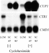
Expression of CUP1 and CTR1 in cells cultured in the presence or absence of cycloheximide. Cells were cultured overnight in LCM (containing 0.1 mM BCS) and incubated in the presence or absence of 0.1 mM cycloheximide for 30 min prior to a medium transition to SC plus 0.3 mM CuSO4. Cycloheximide was also added to the medium after the transition. Cells were harvested 45 or 90 min later and S1 analysis was carried out on extracted RNA.
FIG. 3.
FIG. 3.
Functionality of epitope-tagged Mac1 variants in steady state cultures. Panel A. Expression of CTR1 in wild-type cells, cells containing chromosomally TAP-tagged Mac1 (W303) or mac1Δ BY4741 cells transformed with vectors containing either MAC1 or Myc-tagged MAC1 were evaluated by S1 nuclease analysis. Cells were cultured in LCM, SC, or SC plus 0.3 mM CuSO4 for 7 h. Panel B. Western analysis of tagged Mac1 in steady-state cultures in either LCM, SC, or SC plus 0.3 mM CuSO4 for 7 h. Antisera to Myc, Pgk1, or IgG (for TAP detection). TAP-tagged cells are shown on top and Myc-tagged Mac1 cells are shown on the bottom.
FIG. 4.
FIG. 4.
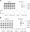
Chromatin immunoprecipitation studies on cells with epitope-tagged Mac1 variants. Panel A. Chromatin immunoprecipitation analysis on steady-state cultures containing either MAC1 or MAC1-Myc. Steady-state cells were cultured in LCM, SC, or SC plus 0.3 mM CuSO4 for 7 h. Cells were harvested at that time and cross-linked for chromatin immunoprecipitation as described in Materials and Methods. Panel B. Chromatin immunoprecipitation analysis on transition cultures containing either MAC1 or MAC1-Myc. Cells grown in LCM were shifted to SC medium and harvested 15 or 30 min later. Binding to Mac1-target gene promoters CTR1, CTR3, and FRE1 as well as the control ZRT1 promoter was tested.
FIG. 5.
FIG. 5.
Chromatin immunoprecipitation studies on cells with TAP epitope-tagged Ace1. Panel A. Ace1-mediated expression of CUP1 in cells undergoing a transition from LCM medium to SC medium. Cells were harvested either 10 or 20 min after the transition and RNA was extracted for S1 nuclease analysis of CUP1 expression levels. Panel B. Chromatin immunoprecipitation analysis on Ace1-TAP cells and control wild-type cells. Cells were grown in LCM and transitioned to SC medium for the times indicated. Binding at the Ace1-target gene CUP1 and the control CTR1 gene was tested.
FIG. 6.
FIG. 6.
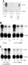
Effects of overexpression of the Ace1 DNA binding domain. Panel A. Wild-type cells with or without ACE1 (DBD) overexpression were cultured in LCM containing raffinose. Galactose was added and 1.5 h later the cells were transitioned to SC medium for 20 min prior to harvest. S1 nuclease analysis was carried out on the harvested cells to assess the effects of overexpression of the Ace1 DBD on expression of CUP1 with CMD1 used as a loading control. Panel B. Cultures grown as in panel A were transitioned to SC medium or SC medium containing 10 μM CuSO4 for 20 min prior to harvest. S1 nuclease analysis was carried out on the harvested cells to assess the effects of overexpression of the Ace1 DBD on expression of CTR1. Panel C. Galactose was added to cells cultured overnight in raffinose SC medium (prepared with BIO 101 low-copper nitrogen base) to induce expression of the ACE1 DBD. Cells were harvested at 0, 45 or 90 min after the addition of galactose. RNA was extracted for S1 nuclease analysis to quantify CTR1 expression levels. Wild-type control cells were cultured in raffinose SC medium in the presence of 0.1 mM BCS to maximally induce CTR1.
FIG. 7.
FIG. 7.
Immunofluorescence of Mac1-GFP and Ace1-GFP chimeras. Cells transformed with GFP fusions of MAC1 or ACE1 were grown in raffinose-containing medium with either 100 μM BCS or 100 μM CuSO4. When the cells reached an optical density of 1, galactose was added to a final concentration of 2% and the cells were incubated for an additional 2 h. Glucose was then added to a final concentration of 2% and the cells grown for an additional hour. The cultures were fixed by the direct addition of formaldehyde (4% final concentration) for 1 h with gentle shaking. The cells were harvested, resuspended in buffered formaldehyde (4% formaldehyde, 50 mM potassium phosphate pH 7.2, 0.5 mM MgCl2) and incubated overnight at 30°C with shaking. The fixed cells were harvested and washed with PBS three times. Upon the addition of 4′,6′-diamidino-2-phenylindole (DAPI) (50 ng/ml) the cells were incubated at 4°C for 48 h. Aliquots of cells were placed on polylysine-coated slides, mounted with FluorSave (Calbiochem), and sealed with a coverslip for microscopy. The overlay of the DAPI image and the GFP fluorescence is shown for both Ace1 and Mac1.
FIG. 8.
FIG. 8.
Effects of overexpression of MAC1 or a mutant MAC1 containing mutations at codons 16 and 19 resulting in Arg to Lys substitutions. Panel A. mac1Δ cells transformed with either MAC1, mutant MAC1 (m) containing the R>K substitutions, or a vector control (vec) were cultured overnight in either LCM or SC plus 10 μM CuSO4 with raffinose as the carbon source. Galactose was added to the cultures and cells were harvested 7 h later. RNA was extracted and used for S1 nuclease analysis to quantify CTR1 expression. Panel B. The same cells used in the panel A experiment were cultured overnight in LCM containing raffinose. Galactose was added and 1.5 h later the cells were transitioned to SC medium or SC medium containing 10 μM CuSO4 for 20 min prior to harvest. S1 nuclease analysis was carried out to quantify expression of CUP1 with CMD1 used as a loading control.
FIG. 9.
FIG. 9.
Effects of overexpressing CRS5 or CRS5 containing a NLS (CRS5-NLS). Wild-type cells transformed with either CRS5, CRS5-NLS or a vector control were cultured in SC medium (prepared with BIO 101 low-copper nitrogen base) containing raffinose. Galactose was added to these cultures and cells were harvested at either 0, 45, or 90 min later to assess the effect of overexpression of the CRS5 alleles on expression of CTR1 by S1 nuclease analysis.

References

    1. Ausubel, F. M., R. Brent, R. E. Kingston, D. D. Moore, J. G. Seidman, J. A. Smith, and K. Struhl (ed.). 1997. Current Protocols in Molecular Biology, vol. 2. John Wiley & Sons, New York, N.Y.
    1. Bloomfield, S. F., G. S. A. B. Stewart, C. E. R. Dodd, I. R. Booth, and E. G. M. Power. 1998. The viable but non-culturable phenomenon explained? Microbiology 144:1-3. - PubMed
    1. Brown, K. R., G. L. Keller, I. J. Pickering, H. H. Harris, G. N. George, and D. R. Winge. 2002. Structures of the cuprous-thiolate clusters of the Mac1 and Ace1 transcriptional activators. Biochemistry 41:6469-6476. - PubMed
    1. Buchman, C., P. Skroch, J. Welch, S. Fogel, and M. Karin. 1989. The CUP2 gene product, regulator of yeast metallothionein expression, is a copper-activated DNA-binding protein. Mol. Cell. Biol. 9:4091-4095. - PMC - PubMed
    1. Carroll, M. C., J. B. Girouard, J. L. Ulloa, J. R. Subramaniam, P. C. Wong, J. S. Valentine, and V. C. Culotta. 2004. Mechanisms for activating Cu- and Zn-containing superoxide dismutase in the absence of the CCS Cu chaperone. Proc. Natl. Acad. Sci. USA 101:5964-5969. - PMC - PubMed

Publication types

MeSH terms

LinkOut - more resources