. 2005 Nov;4(11):1902-12.
doi: 10.1128/EC.4.11.1902-1912.2005.

A chitin synthase and its regulator protein are critical for chitosan production and growth of the fungal pathogen Cryptococcus neoformans

Affiliations

A chitin synthase and its regulator protein are critical for chitosan production and growth of the fungal pathogen Cryptococcus neoformans

Isaac R Banks et al. Eukaryot Cell. 2005 Nov.

Abstract

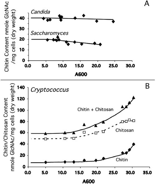
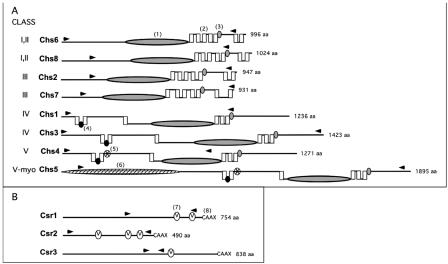
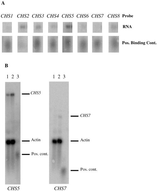
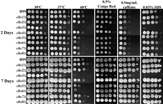
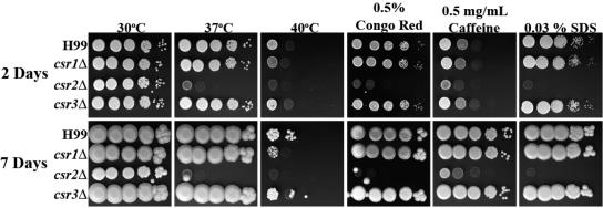
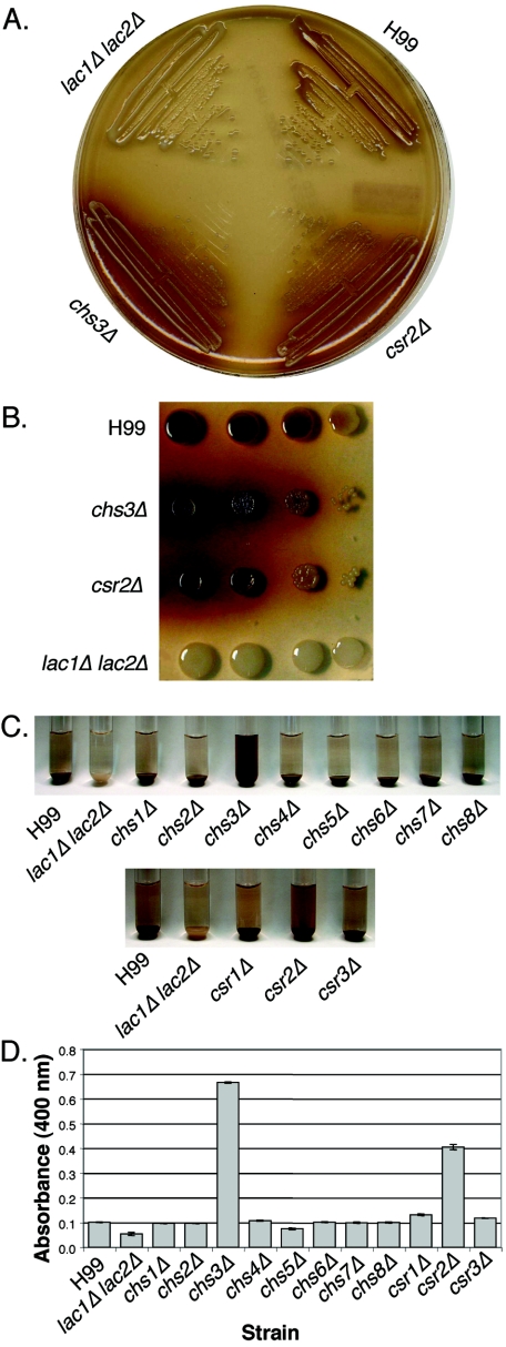
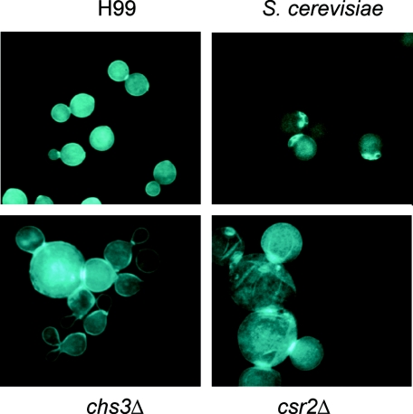
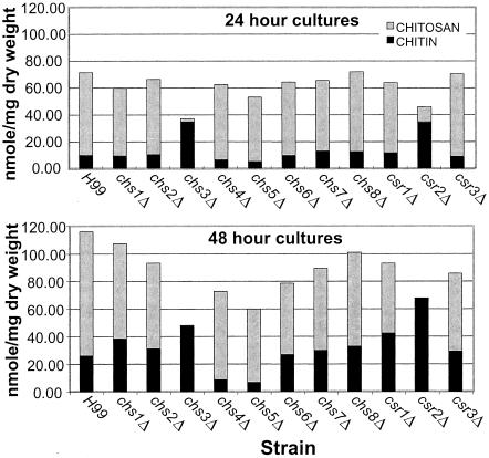
Chitin is an essential component of the cell wall of many fungi. Chitin also can be enzymatically deacetylated to chitosan, a more flexible and soluble polymer. Cryptococcus neoformans is a fungal pathogen that causes cryptococcal meningoencephalitis, particularly in immunocompromised patients. In this work, we show that both chitin and chitosan are present in the cell wall of vegetatively growing C. neoformans yeast cells and that the levels of both rise dramatically as cells grow to higher density in liquid culture. C. neoformans has eight putative chitin synthases, and strains with any one chitin synthase deleted are viable at 30 degrees C. In addition, C. neoformans genes encode three putative regulator proteins, which are homologs of Saccharomyces cerevisiae Skt5p. None of these three is essential for viability. However, one of the chitin synthases (Chs3) and one of the regulators (Csr2) are important for growth. Cells with deletions in either CHS3 or CSR2 have several shared phenotypes, including sensitivity to growth at 37 degrees C. The similarity of their phenotypes also suggests that Csr2 specifically regulates chitin synthesis by Chs3. Lastly, both chs3Delta and the csr2Delta mutants are defective in chitosan production, predicting that Chs3-Csr2 complex with chitin deacetylases for conversion of chitin to chitosan. These data suggest that chitin synthesis could be an excellent antifungal target.


Figures

FIG. 1.
FIG. 1.
Chitin and chitosan measurements of vegetatively growing wild-type C. neoformans strain H99, S. cerevisiae strain BY4741, and C. albicans strain CAI4. YPD cultures were grown and assayed for chitin and chitosan content when cultures reached various absorbance values. Absorbance measurement at 600 nm is indicated at the bottom of the panel. Chitin and chitosan content are indicated at the left of the panel. A. Chitin content of both S. cerevisiae and C. albicans remains virtually constant throughout growth, with no chitosan found for either species during vegetative growth (data not shown). B. Both chitin and chitosan are synthesized by C. neoformans during vegetative growth, the levels of each increasing as culture density increases.
FIG. 2.
FIG. 2.
C. neoformans chitin synthase (Chs) and chitin synthase regulator (Csr) predicted protein structure comparison. The name of each protein is indicated to the immediate left of the protein structure. The predicted amino acid size (from the JEC21 genome sequence) is indicated to the right of each protein structure. A. Chitin synthases. Class designations are indicated by Roman numerals. Chs6 and Chs8 have protein characteristics of both class I and II. Chs5 (class V-myo) has class V protein sequence characteristics as well as a myosin motor domain. Domains were identified by rpsblast searches of the Conserved Domain Database at NCBI (www.ncbi.nlm.nih.gov/Structure/cdd/wrpsb.cgi). Domains are identified as follows: (1) large shaded ellipse, catalytic domain contains homologous sequence of chitin synthase domains 1 and 2 (pfam01644 and pfam03142); (2) vertical box, trans-membrane span; (3) small, shaded oval, conserved sequence “S/T-W-G-X-T/K-R/G”; (4) small black oval, conserved sequence “AWREK”; (5) small starred oval, cytochrome b5-like heme/steroid binding domain (pfam00173); (6) large hatched ellipse, myosin motor domain (cd 00124). B. Chitin synthase regulators. Domains are identified as (7) small “V” oval, SEL1 domain (smart 00671), which may be sites for protein-protein interaction; (8) C-terminal sequence “CAAX” as potential site for farensylation. The area between the arrow heads for each protein in both A and B indicates the region deleted in each deletion strain.
FIG. 3.
FIG. 3.
Chitin synthase expression analysis. A. Relative chitin synthase mRNA expression levels in vegetatively growing H99 (wild type) cells. The top row of bands is the same quantity of H99 RNA probed with each of the chitin synthase genes (labeled at the top). The second row of bands consists of equal amounts of positive binding controls for each chitin synthase (see Materials and Methods). These results are representative of two independent experiments. B. Northern blots showing the increase in transcription of two of the chitin synthase genes in a chs3Δ mutant. Lane 1 is H99 RNA, lane 2 is chs3Δ RNA, and lane 3 is the positive binding control ladder. The blot on the left was probed with CHS5 and actin, and the blot on the right was probed with CHS7 and actin. The dashes on the left indicate where the different transcripts and controls migrate. Pos., positive; cont., control.
FIG. 4.
FIG. 4.
Analysis of H99 and chitin synthase deletion strains for in vitro sensitivity to cell wall stressors. Strains were grown in YPD medium to mid-log phase, serially diluted 10-fold (starting with 1 × 10⁵ cells in the left column of each panel), and plated on YPD solid medium and YPD solid medium supplemented with various cell wall stressors. Strains plated on YPD medium were also tested for high-temperature sensitivity. Time of incubation is indicated at the far left. Strains are indicated at the left of the panels. Incubation conditions are indicated at the top of the panels. All plates containing cell wall stressors were incubated at 30°C.
FIG. 5.
FIG. 5.
Analysis of H99 and chitin synthase regulator deletion strains for in vitro sensitivity to cell wall stressors. The experiment was done as described for Fig. 4.
FIG. 6.
FIG. 6.
Melanin production of H99, chitin synthase deletion strains, and chitin synthase regulator deletion strains. A lac1Δ lac2Δ strain was used as a negative control. A. Cells from chs3Δ and csr2Δ strains were streaked out onto bird seed agar medium and allowed to grow at 30°C for 3 days. B. Serial dilutions of cells spotted onto L-DOPA medium. The spots on the left represent 104 cells, and 10-fold serial dilutions were plated. The strain is indicated to the left of the plate. C. Cells were resuspended to identical optical density in glucose-free asparagine medium containing L-DOPA and allowed to shake at 30°C. Cells were centrifuged to visualize the secreted pigment and the color of the pellets. Pictures were taken at 24 h. The strain name is indicated at the bottom of the panel. D. Quantitation of the OD400 of the supernatants from the cultures as shown in panel C.
FIG. 7.
FIG. 7.
Calcofluor white staining of S. cerevisiae, C. neoformans H99, and deletion strains. Cells were grown in YPD at 30°C, formaldehyde fixed, and stained with calcofluor white, a dye that binds to chitin. The strain is indicated at the left of each photo. All photos are at the same magnification (1,000×).
FIG. 8.
FIG. 8.
Chitin and chitosan content of Cryptococcus neoformans H99 and mutant strains. Measurements were taken at 24-h (top panel) and 48-h (bottom panel) time points. The strains are indicated at the bottom of each graph, and the time of growth is indicated at the top of each graph. Chitin and chitosan content are indicated at the left of each panel.

References

    1. Alspaugh, J. A., R. C. Davidson, and J. Heitman. 2000. Morphogenesis of Cryptococcus neoformans. Contrib. Microbiol. 5:217-238. - PubMed
    1. Araki, Y., and E. Ito. 1975. A pathway of chitosan formation in Mucor rouxii. Enzymatic deacetylation of chitin. Eur. J. Biochem. 55:71-78. - PubMed
    1. Bartnicki-Garcia, S., and E. Lippman.1969. Fungal morphogenesis: cell wall construction in Mucor rouxii. Science 165:302-304. - PubMed
    1. Bartnicki-Garcia, S., and W. J. Nickerson. 1962. Isolation, composition, and structure of cell walls of filamentous and yeast-like forms of Mucor rouxii. Biochim. Biophys. Acta 58:102-119. - PubMed
    1. Bowen, A. R., J. L. Chen-Wu, M. Momany, R. Young, P. J. Szaniszlo, and P. W. Robbins. 1992. Classification of fungal chitin synthases. Proc. Natl. Acad. Sci. USA 89:519-523. - PMC - PubMed

Publication types

LinkOut - more resources