

Spread of human cancer cells occurs with probabilities indicative of a nongenetic mechanism
- PMID: 16278668
- PMCID: PMC2361524
- DOI: 10.1038/sj.bjc.6602848
Spread of human cancer cells occurs with probabilities indicative of a nongenetic mechanism
Abstract
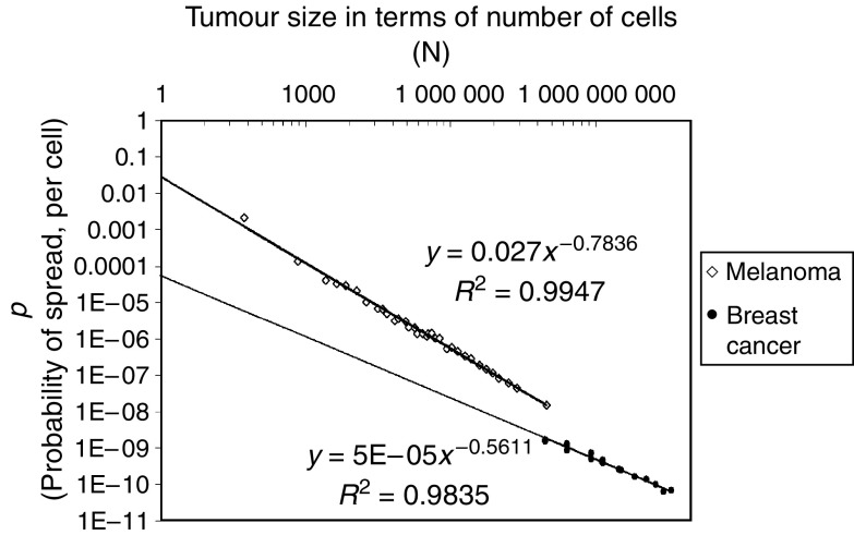
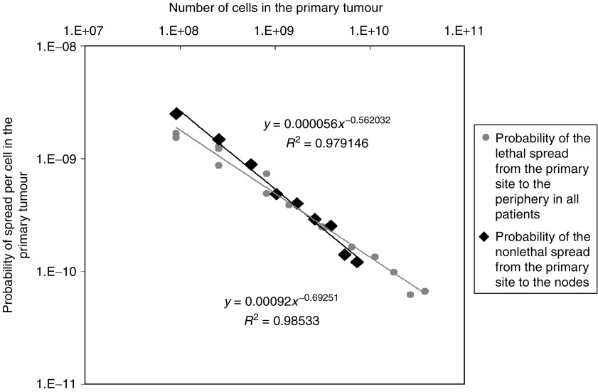
There has been much uncertainty as to whether metastasis requires mutation at the time of spread. Here, we use clinical data to calculate the probability of the spread of melanoma and breast cancer cells. These calculations reveal that the probability of the spread of cancer cells is relatively high for small tumours (approximately 1 event of spread for every 500 cells for melanomas of 0.1 mm) and declines as tumours increase in size (approximately 1 event of spread for every 10(8) cells for melanomas of 12 mm). The probability of spread of breast cancer cells from the lymph nodes to the periphery is approximately 1 event of spread for every 10(8) cells in the nodal masses, which have a mean diameter of 5 mm, while the probability of spread of cancer cells from the breast to the periphery when the primary masses are 5 mm is also approximately 1 event of spread for every 10(8) cells. Thus, the occurrence of an event of spread from the breast to the lymph nodes appears not to increase the propensity of the progeny of those cells to spread from the lymph nodes to the periphery. These values indicate that the spread of human breast cancer and melanoma cells is unlikely to occur by a mechanism requiring mutation at the time of spread.
Figures
References
-
- Balch CM, Soong SJ, Gershenwald JE, Thompson JF, Reintgen DS, Cascinelli N, Urist M, McMasters KM, Ross MI, Kirkwood JM, Atkins MB, Thompson JA, Coit DG, Byrd D, Desmond R, Zhang Y, Liu PY, Lyman GH, Morabito A (2001) Prognostic factors analysis of 17,600 melanoma patients: validation of the American Joint Committee on Cancer melanoma staging system. J Clin Oncol 19: 3622–3634 - PubMed
-
- Bernards R (2003) Clues for migration. Nature 425: 247–248 - PubMed
-
- Bernards R, Weinberg RA (2002) A progression puzzle. Nature 418: 823. - PubMed
-
- Boon ME, Trott PA, van Kaam H, Kurver PJ, Leach A, Baak JP (1982) Morphometry and cytodiagnosis of breast lesions. Virchows Arch [Pathol Anat] 396: 9–18 - PubMed
MeSH terms
LinkOut - more resources
Full Text Sources
Other Literature Sources
Medical
