

doi: 10.1080/08998280.2003.11927882.
Evaluation and management of the incidental adrenal mass
Affiliations
- PMID: 16278716
- PMCID: PMC1200803
- DOI: 10.1080/08998280.2003.11927882
Item in Clipboard
Evaluation and management of the incidental adrenal mass
Proc (Bayl Univ Med Cent).
2003 Jan.
No abstract available
Figures
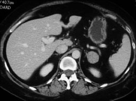
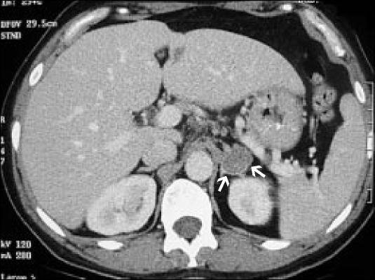
A characteristic incidentaloma. This is an encapsulated solid lesion in the left adrenal gland. Final diagnosis cannot be made without further workup.
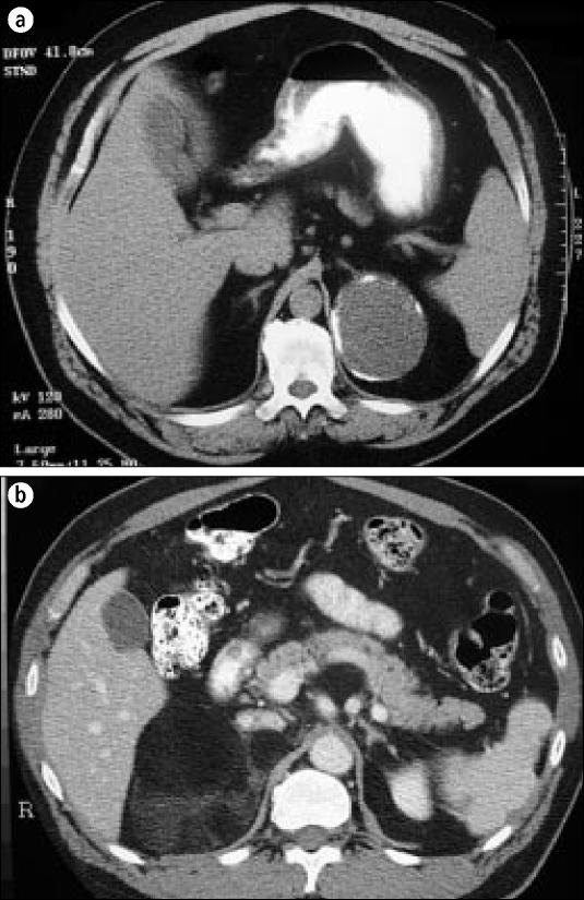
(a) A left adrenal cyst with a fluid-filled cavity and calcified rim. (b) A right myelolipoma with predominantly lipid density.
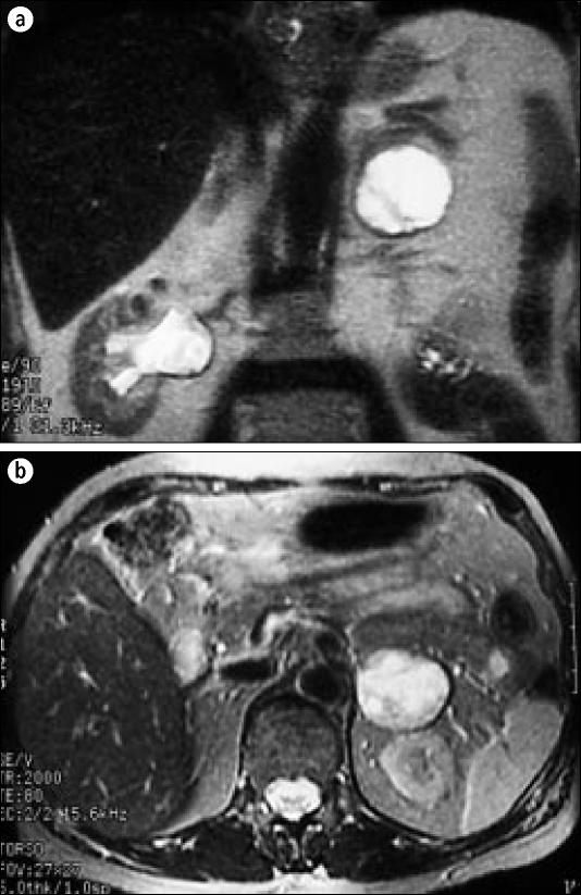
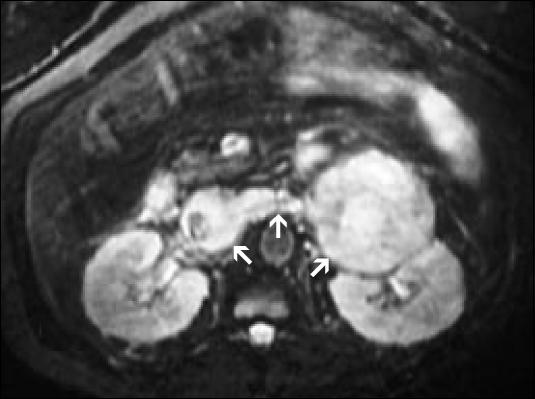
A characteristic (a) coronal and (b) axial MRI T2-weighted sequence of a pheochromocytoma. The left adrenal mass is very bright and easy to identify.
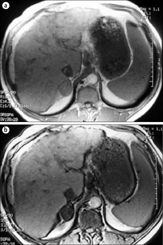
In-phase and (b) opposed-phase chemical-shift MRI. On the opposed-phase chemical shift, the right adenoma darkens—a finding 100% specific for a benign adenoma.
A typical well-encapsulated aldosteronoma of the left adrenal gland. It is benign but is producing aldosterone.
A left adrenocortical carcinoma with renal vein and inferior vena cava invasion on a T2-weighted MRI.
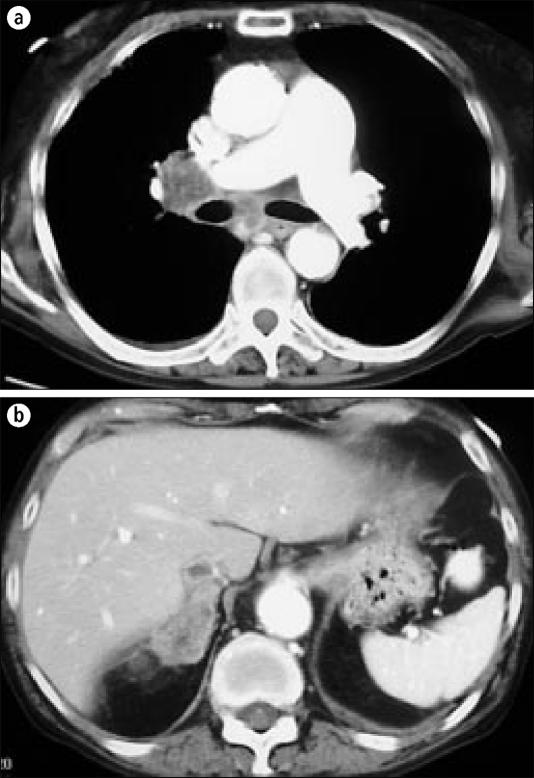
(a) Chest CT scan with right hilar and mediastinal lymphadenopathy in a patient with squamous cell lung cancer, (b) Images through the abdomen demonstrate a right adrenal mass, proven to be a squamous cell cancer metastasis.
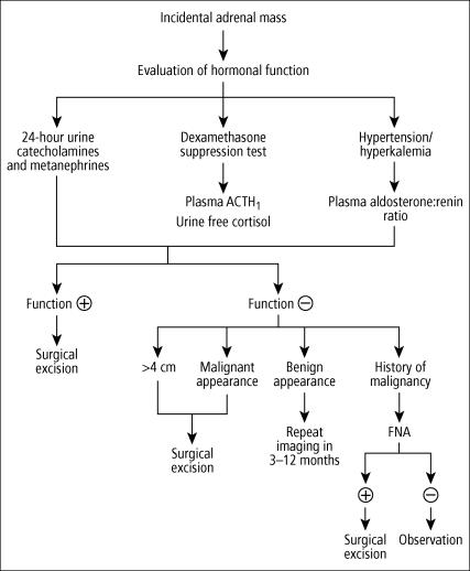
Algorithm for managing adrenal incidentalomas. ACTH indicates adreno-corticotropic hormone; FNA, fine-needle aspiration.
References
-
- Virkkala A, Valimaki M, Pelkonen R, Huikuri K, Kahri A, Kiuisaari L, Korhonen T, Salmi J, Seppala P. Endocrine abnormalities in patients with adrenal tumours incidentally discovered on computed tomography. Acta Endrocrinol (Copenh) 1989;121:67–72. - PubMed
-
- Saruta T. Adrenal incidentaloma Presented at the Ninth International Congress of Endocrinology, Nice, France, August 30, 1992.
-
- Cook DM. Adrenal mass. Endocrinol Metab Clin North Am. 1997;26:829–852. - PubMed
-
- Mitchell DG, Crovello M, Matteucci CT, Petersen RO, Miettinen MM. Benign adrenocortical masses: diagnosis with chemical shift MR imaging. Radiology. 1992;185:345–351. - PubMed
-
- Korobkin M, Francis IR. Imaging of adrenal masses. Urol Clin North Am. 1997;24:603–622. - PubMed
LinkOut - more resources
Full Text Sources