. 2005 Dec;2(12):e355.
doi: 10.1371/journal.pmed.0020355. Epub 2005 Nov 15.

Persistent amyloidosis following suppression of Abeta production in a transgenic model of Alzheimer disease

Affiliations

Persistent amyloidosis following suppression of Abeta production in a transgenic model of Alzheimer disease

Joanna L Jankowsky et al. PLoS Med. 2005 Dec.

Abstract

Background: The proteases (secretases) that cleave amyloid-beta (Abeta) peptide from the amyloid precursor protein (APP) have been the focus of considerable investigation in the development of treatments for Alzheimer disease. The prediction has been that reducing Abeta production in the brain, even after the onset of clinical symptoms and the development of associated pathology, will facilitate the repair of damaged tissue and removal of amyloid lesions. However, no long-term studies using animal models of amyloid pathology have yet been performed to test this hypothesis.

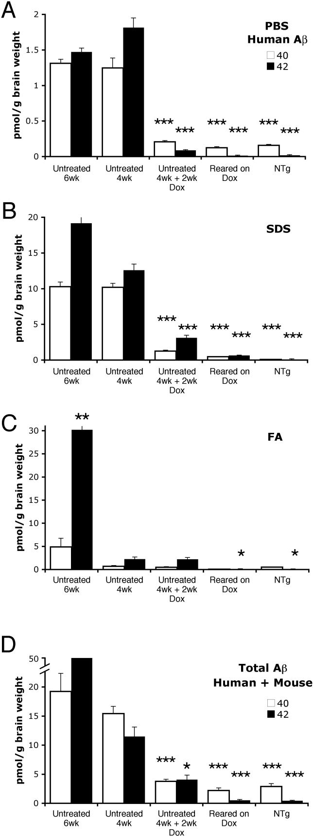
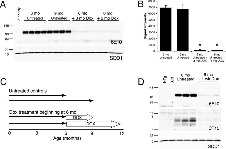
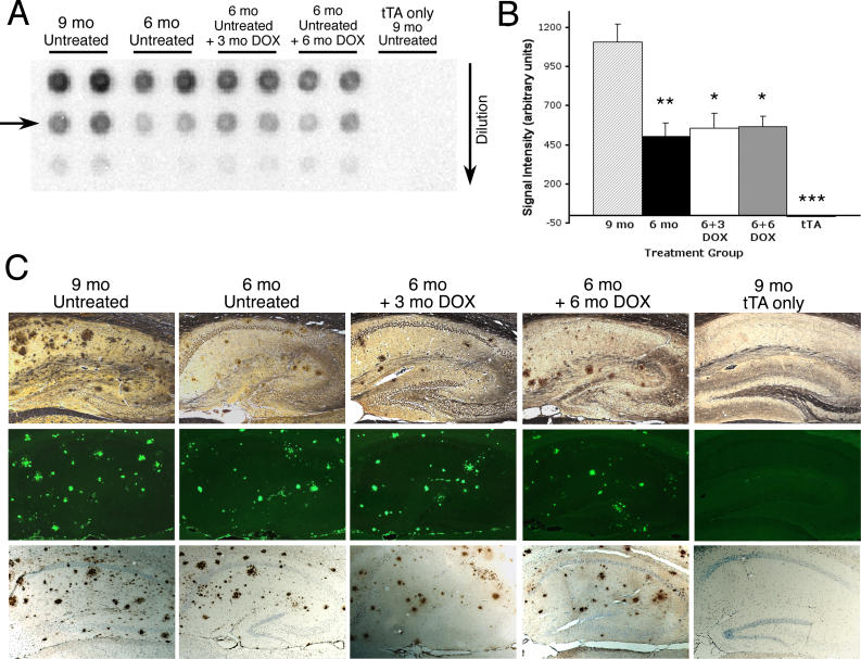
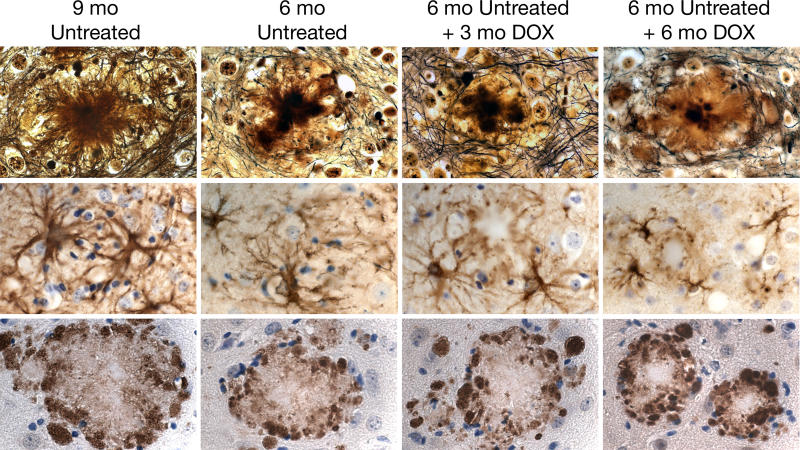
Methods and findings: We have generated a transgenic mouse model that genetically mimics the arrest of Abeta production expected from treatment with secretase inhibitors. These mice overexpress mutant APP from a vector that can be regulated by doxycycline. Under normal conditions, high-level expression of APP quickly induces fulminant amyloid pathology. We show that doxycycline administration inhibits transgenic APP expression by greater than 95% and reduces Abeta production to levels found in nontransgenic mice. Suppression of transgenic Abeta synthesis in this model abruptly halts the progression of amyloid pathology. However, formation and disaggregation of amyloid deposits appear to be in disequilibrium as the plaques require far longer to disperse than to assemble. Mice in which APP synthesis was suppressed for as long as 6 mo after the formation of Abeta deposits retain a considerable amyloid load, with little sign of active clearance.

Conclusion: This study demonstrates that amyloid lesions in transgenic mice are highly stable structures in vivo that are slow to disaggregate. Our findings suggest that arresting Abeta production in patients with Alzheimer disease should halt the progression of pathology, but that early treatment may be imperative, as it appears that amyloid deposits, once formed, will require additional intervention to clear.


Conflict of interest statement

Competing Interests: The authors have declared that no competing interests exist.

Figures

Figure 1
Figure 1. Control of Transgenic APP Expression by Dox
(A) Western blotting for transgenic APP using the human-specific 6E10 antibody shows expression of full-length protein in forebrain tissue from young predeposit double transgenic animals (line 107) and its suppression following dox treatment. Untreated animals show high levels of transgene expression; protein levels drop dramatically in animals acutely treated with dox for 2 wk. A faint, but detectable band of full-length protein remains in acutely treated animals that can be eliminated in mice born and raised on dox. (B) Immunoblotting with the N-terminal antibody 22C11 to detect both transgenic and endogenous protein shows that dox treatment reduces APP/APLP to levels found in nontransgenic mice. (C) Measurement of signal intensity from the Western blot in (A) shows transgenic APP levels are decreased more than 95% by dox in both acutely and chronically treated animals (97.2% for 4 wk + 2 wk dox, 98.0% for reared on dox versus 4 wk untreated; ANOVA effect of treatment group F 4,15 = 85.55, p < 0.001). APP levels in 4 wk + 2 wk dox, reared on dox, and nontransgenic (NTg) were not significantly different (p > 0.9, Tukey post-hoc test). (D) Measurement of signal intensity from the Western blot in (B) shows total APP/APLP levels in dox-treated tTA/APP mice are significantly lower than in 4-wk-old untreated mice (ANOVA effect of treatment group F 4,15 = 84.41, p < 0.001) and indistinguishable from those of nontransgenic animals (p > 0.9, Tukey post-hoc test). *, p < 0.001 versus untreated 4-wk-old mice, Tukey post-hoc test applied to significant effect of group ANOVA.
Figure 2
Figure 2. Aβ Levels Are Dramatically Reduced by Transgene Suppression
Cortical homogenates from young, predeposit tTA/APP mice used for Western blot in Figure 1 (line 107) were fractionated by sequential multi-step extraction with PBS, 2% SDS, and 70% FA followed by human-specific Aβ ELISA to measure transgene-derived peptide in each fraction. Aβ40 is shown in white, Aβ42 in black. (A) PBS-soluble Aβ levels are substantially reduced by both acute and chronic dox treatment (ANOVA, effect of treatment group F 4,24 = 137.10 and 386.01, p < 0.001, for Aβ40 and Aβ42, respectively). Aβ levels in treated animals are indistinguishable from nontransgenic (NTg) animals (p > 0.3, Tukey post-hoc test). (B) In the young animals tested here prior to the formation of visible amyloid deposits, most Aβ is extracted into the SDS fraction (84% and 76% of all transgene-derived Aβ40 and Aβ42, respectively). As in the PBS-soluble fraction, Aβ levels in the SDS fraction are significantly lowered by dox treatment compared to untreated animals (ANOVA effect of group F 4,24 = 197.57 and 163.48, p < 0.001, for Aβ40 and Aβ42, respectively). Acutely treated animals retained a small (although significant) amount of residual peptide (p < 0.001 compared to nontransgeinc, Tukey post-hoc test), whereas Aβ levels in mice born and raised on dox were reduced to levels indistinguishable from nontransgenic (p > 0.8, Tukey post-hoc test). (C) The FA-soluble fraction already contains a small but significant pool of aggregated Aβ42 in untreated animals by 4 wk of age (p < 0.05 versus nontransgenic; Tukey post-hoc test applied to significant effect of group ANOVA F 4,24 = 17.11, p < 0.001). By 6 wk of age, the amount of Aβ in the FA fraction is increased significantly preceding the appearance of visible deposits 2 wk later. The FA pool is the only peptide fraction not lowered by acute dox treatment (4 wk untreated = 4 wk + 2 wk dox, p > 0.9, Tukey post-hoc test), consistent with poor turnover of aggregated Aβ species. (D) Measurements of total Aβ, including both endogenous and transgene-derived peptides, show that animals born and raised on dox harbor Aβ levels identical to nontransgenic animals (p > 0.9, Tukey post-hoc test, effect of group ANOVA F 4,24 = 39.13 and 35.29, p < 0.001, for Aβ40 and Aβ42, respectively). Whereas chronic transgene suppression fully prevents synthesis of both peptides, acute dox treatment fully suppresses Aβ40 levels (p > 0.8, Tukey post-hoc test), but leaves a small amount of nonsuppressed Aβ42. The residual Aβ42 observed in acutely treated young animals derives from uncleared aggregates extracted in the SDS and FA fractions. *, p < 0.05; **, p < 0.005; ***, p < 0.001 versus 4-wk-old untreated mice, Tukey post-hoc applied to significant effect of group ANOVA.
Figure 3
Figure 3. Robust Transgene Suppression in Older Mice with Preexisting Amyloid Pathology
(A) Cortical homogenates from 6- to 12-mo-old animals used for pathology studies described below (line 107) were immunoblotted with human-specific antibody 6E10 to examine transgene suppression following 3 or 6 mo of dox treatment. The blot was co-immunostained for endogenous superoxide dismutase 1 (SOD1) as a control for loading. (B) Quantitation of signal intensity from the Western blot shown in (A). Transgenic APP levels are significantly suppressed following 3 or 6 mo of dox treatment (96.9% and 97.6%, respectively). *, p < 0.001 compared to 6-mo-old untreated animals, Tukey post-hoc test applied to significant effect of group ANOVA F 3,12 = 107.22, p < 0.001. These data demonstrate that strong transgene suppression is attained both before and after the onset of amyloid pathology (see Figure 1 for predeposit experiments). (C) Experimental design. To examine the effects of chronic Aβ suppression on amyloid pathology after the onset of deposition, we compared untreated controls harvested at 6 and 9 mo of age to animals placed on dox at 6 mo of age and harvested after 3 or 6 mo of treatment. (D) Dox treatment leads to rapid transgene suppression even in 6-mo-old tTA/APP mice. Immunostaining with 6E10 shows APPswe/ind levels are dramatically reduced in 6-mo-old mice treated for 1 wk with dox (upper panel). A separate blot was immunostained for APP C-terminal fragments with CT15 antibody to show that the precursors to Aβ cleavage are decreased in parallel with the full-length protein (middle panel). Costaining for superoxide dismutase 1 was used as an internal control for loading (lower panel, taken from bottom half of 6E10 blot).
Figure 4
Figure 4. Suppression of Transgenic APP Arrests Progression of Amyloid Pathology
(A) Aggregated Aβ was quantified in cortical tissue from dox-treated and control tTA/APP mice (line 107) using a filter trap assay. Serial dilutions of protein homogenate were passed through a cellulose acetate filter; protein aggregates larger than the pore size were trapped and immunostained for Aβ. (B) Quantitation of signal intensity in the linear range of each filter trap dilution series (arrow in [A]) was used to compare aggregate load across treatment groups. Aggregated Aβ increased significantly between 6 and 9 mo of age in untreated mice (significant effect of group ANOVA F 3,18 = 7.85, p < 0.002). This progression of pathology was completely prevented by transgene suppression. The amount of aggregated Aβ was identical in untreated mice at 6 mo of age to that in 9- or 12-mo-old animals treated with dox (p > 0.9, Tukey post-hoc test). Single transgenic tTA samples were included as negative controls and showed no signal above background. *, p < 0.01; **, p < 0.005 versus 9-mo-old untreated mice, Tukey post-hoc test; ***, p < 0.001 versus 9-mo-old untreated mice, Student's t-test. (C) Amyloid pathology in the hippocampus of representative mice from each treatment group: Hirano silver stain (top row), thioflavin-S (middle row), and Aβ immunohistochemistry (bottom row). Amyloid burden increases dramatically between 6 and 9 mo of age in untreated animals, but remains stable in transgene-suppressed mice over the same period (6 mo + 3 mo dox and 6 mo + 6 mo dox). Single transgenic animals (tTA only shown here) show no sign of amyloid pathology at any age tested.
Figure 5
Figure 5. Aβ ELISA Confirms Arrest of Progression without Clearance of Peptide in Mice with Preexisting Aggregates
Aβ levels in untreated 6- and 9-mo-old tTA/APP line 107 mice (shown in Figure 4) were compared to those in 9- and 12-mo-old animals treated with dox from the age of 6 mo. Single transgenic APP samples were included as negative controls. Cortical homogenates were fractionated by sequential multi-step extraction with PBS, 2% SDS, and 70% FA followed by human-specific Aβ ELISA to measure transgene-derived peptide in each fraction. Aβ40 is shown in white, Aβ42 in black. (A and B) Most Aβ in the brains of plaque-bearing mice is extracted into the FA and SDS fractions. Consistent with amyloid burden (Figures 4 and Figure S3), SDS- and FA-extracted Aβ levels in untreated 9-mo-old mice were significantly higher than in untreated 6-mo-old mice (Tukey post-hoc test applied to significant effect of group ANOVA for SDS and FA fractions F 3,18 = 4.72–12.92, p < 0.02). In contrast, 3 or 6 mo of transgene suppression held Aβ at levels equivalent to those harbored when treatment was started (p > 0.2 compared to 6 mo untreated mice, Tukey post-hoc test). *, p < 0.05; **, p < 0.005; ***, p < 0.001 versus 9-mo-old untreated mice, Tukey post-hoc test. Significance for APP versus 9-mo-old untreated mice is based on Student's t-test. (C) The PBS fraction represents less than 0.1% of total Aβ (note the change in y-axis from [A] and [B]), but only here do Aβ levels in the dox-treated mice differ from those in younger untreated mice. Although both peptides appear elevated in the treated groups compared to the untreated 6-mo-old mice, only Aβ40 reaches statistical significance (p < 0.05, Tukey post-hoc test applied to significant effect of group ANOVA for Aβ40 F 3,18 = 4.60, p < 0.02). A similar trend was seen for Aβ42, where ANOVA yielded a significant effect of group for PBS-soluble Aβ42 (F 3,18 = 3.75, p < 0.03), however this was due only to differences between the untreated 6- and 9-mo-old groups. •, p < 0.05 versus 6-mo-old untreated mice, Tukey post-hoc test; •••, p < 0.001 versus 6-mo-old untreated mice, Student's t-test.
Figure 6
Figure 6. Neuritic and Glial Pathology Are Unchanged following Transgene Suppression
Dystrophic neurites and activated astrocytes surround most compact plaques in tet-off APP mice (line 107). Dark-stained, ubiquitin-filled neurites and reactive astrocytes form a halo around cored, fibrillar deposits by 6 mo of age that worsens with time in untreated mice. Both plaque-associated pathologies are arrested, although not reversed, by transgene suppression. Hirano silver stain (top row); GFAP immunohistochemistry (middle row); ubiquitin immunohistochemistry (bottom row).
Figure 7
Figure 7. Transgene Suppression Attenuates Hyperactivity in tTA/APP Mice
(A) A 48-h measure of ambulation records extreme hyperactivity in untreated double transgenic mice compared to single transgenic and nontransgenic controls (line 107). This phenotype is completely eliminated by rearing the double transgenic mice on dox. (B) The same data shown in (A) are replotted to magnify data from untreated control and dox-treated groups. (C and D) Activity levels in the combined control groups of (A) and (B) are here separated by genotype. None of the single transgenic or nontransgenic control groups display the hyperactivity present in untreated tTA/APP animals. Again, note the y-axes have been enlarged for detail compared to (A).

Comment in

  • Breaking up (amyloid) is hard to do.
    Gandy S, Heppner FL. Gandy S, et al. PLoS Med. 2005 Dec;2(12):e417. doi: 10.1371/journal.pmed.0020417. Epub 2005 Dec 27. PLoS Med. 2005. PMID: 16363913 Free PMC article.

References

    1. Hardy JA, Higgins GA. Alzheimer's disease: The amyloid cascade hypothesis. Science. 1992;256:184–185. - PubMed
    1. Selkoe DJ, Podlisny MB. Deciphering the genetic basis of Alzheimer's disease. Annu Rev Genomics Hum Genet. 2002;3:67–99. - PubMed
    1. Rademakers R, Cruts M, Van Broeckhoven C. Genetics of early-onset Alzheimer dementia. ScientificWorldJournal. 2003;3:497–519. - PMC - PubMed
    1. Orgogozo JM, Gilman S, Dartigues JF, Laurent B, Puel M, et al. Subacute meningoencephalitis in a subset of patients with AD after Aβ42 immunization. Neurology. 2003;61:46–54. - PubMed
    1. Check E. Nerve inflammation halts trial for Alzheimer's drug. Nature. 2002;415:462. - PubMed

Publication types

Substances