. 2005 Dec;2(12):e381.
doi: 10.1371/journal.pmed.0020381. Epub 2005 Nov 15.

DC-SIGN induction in alveolar macrophages defines privileged target host cells for mycobacteria in patients with tuberculosis

Affiliations

DC-SIGN induction in alveolar macrophages defines privileged target host cells for mycobacteria in patients with tuberculosis

Ludovic Tailleux et al. PLoS Med. 2005 Dec.

Abstract

Background: Interplays between Mycobacterium tuberculosis, the etiological agent of tuberculosis (TB) in human and host professional phagocytes, namely macrophages (Mphis) and dendritic cells (DCs), are central to immune protection against TB and to TB pathogenesis. We and others have recently shown that the C-type lectin dendritic cell-specific intercellular adhesion molecule-3 grabbing nonintegrin (DC-SIGN; CD209) mediates important interactions between mycobacteria and human monocyte-derived DCs (MoDCs) in vitro.

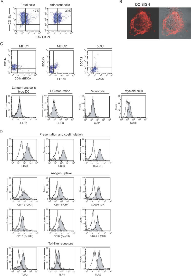
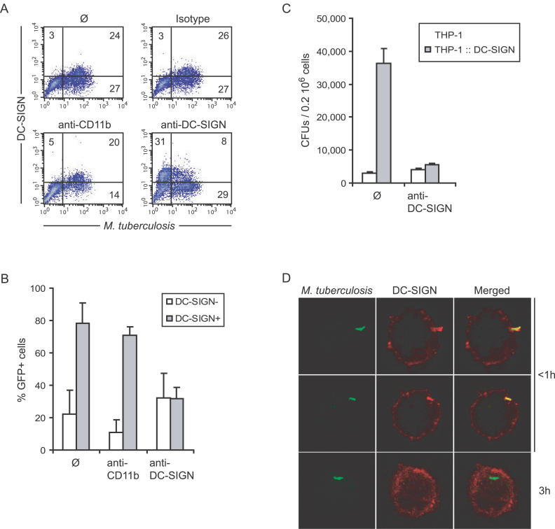
Methods and findings: In order to explore the possible role of DC-SIGN in M. tuberculosis infection in vivo, we have analysed DC-SIGN expression in broncho-alveolar lavage (BAL) cells from patients with TB (n = 40) or with other non-mycobacterial lung pathologies, namely asthma (n = 14) and sarcoidosis (n = 11), as well as from control individuals (n = 9). We show that in patients with TB, up to 70% of alveolar Mphis express DC-SIGN. By contrast, the lectin is barely detected in alveolar Mphis from all other individuals. Flow cytometry, RT-PCR, and enzyme-linked immunosorbent assay analyses of BAL-derived fluids and cells indicated that M. tuberculosis infection induces DC-SIGN expression in alveolar Mphis by a mechanism that is independent of Toll-like receptor-4, interleukin (IL)-4, and IL-13. This mechanism most likely relies on the secretion of soluble host and/or mycobacterial factors that have yet to be identified, as both infected and uninfected bystander Mphis were found to express DC-SIGN in the presence of M. tuberculosis. Immunohistochemical examination of lung biopsy samples from patients with TB showed that the bacilli concentrate in pulmonary regions enriched in DC-SIGN-expressing alveolar Mphis in vivo. Ex vivo binding and inhibition of binding experiments further revealed that DC-SIGN-expressing alveolar Mphis constitute preferential target cells for M. tuberculosis, as compared to their DC-SIGN- counterparts. In contrast with what has been reported previously in MoDCs in vitro, ex vivo DC-SIGN ligation by mycobacterial products failed to induce IL-10 secretion by alveolar Mphis, and IL-10 was not detected in BALs from patients with TB.

Conclusion: Altogether, our results provide further evidence for an important role of DC-SIGN during TB in humans. DC-SIGN induction in alveolar Mphis may have important consequences on lung colonization by the tubercle bacillus, and on pulmonary inflammatory and immune responses in the infected host.


Conflict of interest statement

Competing Interests: The authors have declared that no competing interests exist.

Figures

Figure 1
Figure 1. Alveolar CD11b+ Cells Over-Express DC-SIGN in Patients with TB
(A) BAL cells from a patient with TB (upper four panels) and from a patient with sarcoidosis (lower two panels) were analyzed by flow cytometry. Expression of CD3 and CD4 was analyzed on cells from R1. CD11b and DC-SIGN expression was analyzed on cells from R2. (B) Distribution of the proportion of CD11b+DC-SIGN+ cells in BALs according to pathology and age. Black circles indicate ≤15 y of age; black triangles indicate ≥20 y; NC, no case. (C) DC-SIGN (upper panels) and M. tuberculosis (lower panels) immunodetection in serial sections of a lung biopsy from a patient with TB. The pictures are representative of results obtained with samples from a total of four patients. G, granuloma. (D) DC-SIGN immunodetection in a lung biopsy from a patient with sarcoidosis. The pictures are representative of results obtained with samples from a total of three patients. In (C) and (D), magnification in left panels is 100×, and regions in squares are shown at higher magnification in right panels.
Figure 2
Figure 2. Alveolar DC-SIGN+ Cells in Patients with TB Are Mφs
(A) Total BAL cells from a patient with TB were allowed to adhere to the plastic for 1 h at 37 °C in complete medium. CD11b and DC-SIGN expression was analyzed by flow cytometry before (left) and after (right) adherence. (B) Surface and intracellular DC-SIGN (red) expression by an adherent alveolar cell examined under the confocal microscope. (C) Flow cytometry analysis of surface expression of BDCA-1 (CD1c), BDCA-2, BDAC-3, CD1a, CD11b, CD11c, CD14, CD68, CD83, and CD123 in DC-SIGN+ BAL cells from a patient with TB. (D) Flow cytometry analysis of surface expression of CD40, CD86, HLA-DR, CD11b, CD11c, CD206, CD16, CD32, CD40, CD64, TLR2, TLR4, and TLR9 in DC-SIGN+ BAL cells from a patient with TB. In (C) and (D), analysis was performed on DC-SIGN–expressing cells in R2, as shown in Figure 1.
Figure 3
Figure 3. DC-SIGN Is Induced on Resident Alveolar Mφs upon M. tuberculosis Infection
(A) Flow cytometry analysis of CD11b and DC-SIGN expression by PBMCs from a healthy donor (upper panels) and a patient with TB (lower panels). (B) Adherent DC-SIGN alveolar Mφs from a non-tuberculous patient were infected with a GFP-expressing strain of M. tuberculosis at a MOI of one bacterium per cell, or treated with IL-4, TNF-α, or LPS, or left untreated (ø control). After 48 h at 37 °C, cells were recovered and DC-SIGN expression was analyzed by flow cytometry. In M. tuberculosis panel, the grey area corresponds to GFP+ (infected) cells, and the plain line corresponds to GFP (uninfected) cells. (C) Cells infected with M. tuberculosis at a MOI of 1 for 1, 2, 4, or 24 h were analysed by RT-PCR for DC-SIGN and GAPDH mRNAs.
Figure 4
Figure 4. DC-SIGN Mediates M. tuberculosis Binding to Alveolar Mφs from Patients with TB
(A) Alveolar Mφs from a patient with TB were infected with GFP-expressing M. tuberculosis, in the absence (ø; upper left panel) or the presence of control isotype (upper right panel), anti-CD11b (lower left panel), or -DC-SIGN (lower right panel) blocking antibodies. In the upper panels, cells were then stained with fluorescent PE-conjugated anti-DC-SIGN and APC-conjugated anti-CD11b antibodies. In lower panels, fluorescent antibodies were added together with blocking antibodies (same clones). (B) Proportion of GFP+ cells in DC-SIGN (open bars) and DC-SIGN+ (grey bars) alveolar Mφs as calculated from (A) using BALs from two patients with TB. THP1 Mφs expressing or not expressing DC-SIGN (THP1::DC-SIGN) were used in a binding experiment with M. tuberculosis H37Rv, in the presence or absence of anti-DC-SIGN antibodies. (D) Confocal microscopy examination of adherent DC-SIGN+ cells infected with GFP-expressing M. tuberculosis for various times.

References

    1. Flynn JL, Chan J. Immunology of tuberculosis. Annu Rev Immunol. 2001;19:93–129. - PubMed
    1. Kaufmann SH. How can immunology contribute to the control of tuberculosis? Nat Rev Immunol. 2001;1:20–30. - PubMed
    1. Russell DG. Mycobacterium tuberculosis Here today, and here tomorrow. Nat Rev Mol Cell Biol. 2001;2:569–577. - PubMed
    1. Curtis BM, Scharnowske S, Watson AJ. Sequence and expression of a membrane-associated C-type lectin that exhibits CD4-independent binding of human immunodeficiency virus envelope glycoprotein gp120. Proc Natl Acad Sci U S A. 1992;89:8356–8360. - PMC - PubMed
    1. Geijtenbeek TB, Kwon DS, Torensma R, van Vliet SJ, van Duijnhoven GC, et al. DC-SIGN, a dendritic cell-specific HIV-1-binding protein that enhances trans-infection of T cells. Cell. 2000;100:587–597. - PubMed

Publication types

MeSH terms

Substances