. 2005;7(6):R965-74.
doi: 10.1186/bcr1329. Epub 2005 Sep 30.

Rac1 and Rac3 isoform activation is involved in the invasive and metastatic phenotype of human breast cancer cells

Affiliations

Rac1 and Rac3 isoform activation is involved in the invasive and metastatic phenotype of human breast cancer cells

Paige J Baugher et al. Breast Cancer Res. 2005.

Abstract

Introduction: The metastatic progression of cancer is a direct result of the disregulation of numerous cellular signaling pathways, including those associated with adhesion, migration, and invasion. Members of the Rac family of small GTPases are known to act as regulators of actin cytoskeletal structures and strongly influence the cellular processes of integrin-mediated adhesion and migration. Even though hyperactivated Rac proteins have been shown to influence metastatic processes, these proteins have never been directly linked to metastatic progression.

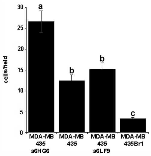
Methods: To investigate a role for Rac and Cdc42 in metastatic breast cancer cell invasion and migration, relative endogenous Rac or Cdc42 activity was determined in a panel of metastatic variants of the MDA-MB-435 metastatic human breast cancer cell line using a p21-binding domain-PAK pull down assay. To investigate the migratory and invasive potential of the Rac isoforms in human breast cancer, namely Rac1 and the subsequently cloned Rac3, we stably expressed either dominant active Rac1 or dominant active Rac3 into the least metastatic cell variant. Dominant negative Rac1 or dominant negative Rac3 were stably expressed in the most metastatic cell variant. Cell lines expressing mutant Rac1 or Rac3 were analyzed using in vitro adhesion, migration and invasion assays.

Results: We show that increased activation of Rac proteins directly correlates with increasing metastatic potential in a panel of cell variants derived from a single metastatic breast cancer cell line (MDA-MB-435). The same correlation could not be found with activated Cdc42. Expression of a dominant active Rac1 or a dominant active Rac3 resulted in a more invasive and motile phenotype. Moreover, expression of either dominant negative Rac1 or dominant negative Rac3 into the most metastatic cell variant resulted in decreased invasive and motile properties.

Conclusion: This study correlates endogenous Rac activity with high metastatic potential and implicates Rac in the regulation of cell migration and invasion in metastatic breast cancer cells. Taken together, these results suggest a role for both the Rac1 and Rac3 GTPases in human breast cancer progression.


Figures

Figure 1
Figure 1
Characterization of cytoskeletal structures and focal adhesion distribution in MDA-MB-435 metastatic variants. (a) Each of the MDA-MB-435 metastatic variants was plated onto glass coverslips. Actin was visualized with rhodamine phalloidin and focal adhesions were visualized with an anti-p-tyro antibody followed by an FITC conjugated secondary antibody. (b) Cell area was measured on 50 individual cells per variant using Spot Digital Camera Software. Focal adhesion number was divided by cell area and plotted on the y-axis. Bars represent ±SEM, and are representative of three independent experiments. Treatments denoted by the same letter indicate no significant difference between those treatments. Treatments denoted by different letters indicate a significant difference between those treatments (P < 0.01).
Figure 2
Figure 2
Haptotaxis assays of MDA-MB-435 metastatic variants. Each variant was adjusted to 500,000 cells and applied to Transwell chambers in a haptotaxis assay, where the underside of the membrane was coated with matrigel. Cells migrating to the underside of the chamber were stained with propidium iodide and counted under 400× Magnification. Bars represent ±SEM. Data are representative of three independent experiments. Treatments denoted by the same letter indicate no significant difference between those treatments. Treatments denoted by different letters indicate a significant difference between those treatments (P < 0.01, calculated from unpaired t-test).
Figure 3
Figure 3
Rac and Cdc42 activity in MDA-MB-435 metastatic variants. Whole cell lysates of all variants were subjected to SDS-PAGE followed by western blot analysis for (a) total Rac using an anti-Rac antibody and (b) total Cdc42 using an anti-Cdc42 antibody. Rac and Cdc42 activities were assayed using the PAK-PBD activity assay. A non-hydrolyzable GTP analog, GTPγS, was used as the positive control; GDP alone was used for the negative control. Equal loading of lanes was maintained by performing a total protein assay and is confirmed by western blot analysis for total actin. Results are representative of three to five independent experiments.
Figure 4
Figure 4
Inhibition of Rho GTPases, Rac, or Cdc42 in migration of the high metastatic MDA-MB-435α6HG6 cell variant. (a) MDA-MB-435α6HG6 cells were treated with vehicle or 2 ng/ml toxin B for 24 h and subjected to a basement membrane haptotaxis assay. Cells migrating to the underside of the membrane were stained with propidium iodide and counted under 400× Magnification. (b) MDA-MB-435α6HG6 cells transiently expressing vector alone or myc-Cdc42(T17N) were subjected to a basement membrane haptotaxis assay. Cells migrating to the underside of the membrane were stained with propidium iodide and counted under 400× Magnification. Equal loading was confirmed by a total actin blot, ectopic myc-Cdc42(T17N) expression confirmed by western blotting with anti-Cdc42 or anti-myc. (c) MDA-MB-435α6HG6 cells transiently expressing vector alone, myc-Rac1(T17N), or myc-Rac3(T17N) were subjected to a basement membrane haptotaxis assay. Bars represent ±SEM; equal loading was confirmed by total actin blot. Myc-Rac1(T17N) and myc-Rac3(T17N) expression were confirmed by western blotting with anti-Rac or anti-myc. Data are expressed as mean ±SEM of three independent experiments. A star denotes statistical significance from control (P < 0.05, calculated from paired t-tests).
Figure 5
Figure 5
Ectopic dominant active Rac(G12V) expression in low metastatic variant MDA-MB-435Br1. Confocal DIC (Differential Interference Contrast) and fluorescent microscopy was performed on MDA-MB-435Br1 cell variant stably expressing vector alone, myc-Rac1(G12V), or myc-Rac3(G12V). Cells were plated on glass coverslips, fixed in 3.7% formaldehyde and permeablized with 0.2% Triton X-100. Actin was then visualized with rhodamine phalloidin and focal adhesions were visualized with an anti-p-tyro antibody followed by an FITC conjugate.
Figure 6
Figure 6
Effects of mutant Rac isoforms on metastatic properties in vitro. MDA-MB-435Br1 cells transiently transfected with vector alone, myc-Rac1(G12V), or myc-Rac3(G12V), were subjected to (a) adhesion, (b) haptotaxis, and (c) invasion assays. MDA-MB-435α6HG6 cells transiently expressing vector alone, myc-Rac1(T17N), or myc-Rac3(T17N), were subjected to (d) adhesion, and (e) invasion assays. Cells were counted under 200× Magnification for adhesions assays, and 400× Magnification for haptotaxis and invasion assays. Y-axis represents the number of cells/field for at least 20 microscopic fields per variant. Bars represent standard error of the mean, and are representative of at least three separate experiments. An asterix indicates a statistically significant difference compared to the control, vector alone, as determined by a paired Student's t-test (P < 0.05).

Similar articles

Cited by

References

    1. Condeelis JS, Wyckoff JB, Bailly M, Pestell R, Lawrence D, Backer J, Segall JE. Lamellipodia in invasion. Semin Cancer Biol. 2001;11:119–128. doi: 10.1006/scbi.2000.0363. - DOI - PubMed
    1. Ridley AJ, Schwartz MA, Burridge K, Firtel RA, Ginsberg MH, Borisy G, Parsons JT, Horwitz AR. Cell migration: integrating signals from front to back. Science. 2003;302:1704–1709. doi: 10.1126/science.1092053. - DOI - PubMed
    1. Price LS, Collard JG. Regulation of the cytoskeleton by Rho-family GTPases: implications for tumour cell invasion. Semin Cancer Biol. 2001;11:167–173. doi: 10.1006/scbi.2000.0367. - DOI - PubMed
    1. Lee TK, Man K, Ho JW, Wang XH, Poon RT, Sun KW, Ng KT, Ng IO, Xu R, Fan ST. The significance of Rac signaling pathway in HCC cell motility: implication for new therapeutic target. Carcinogenesis. 2004;26:681–687. doi: 10.1093/carcin/bgi002. - DOI - PubMed
    1. Bouzahzah B, Albanese C, Ahmed F, Pixley F, Lisanti MP, Segall JD, Condeelis J, Joyce D, Minden A, Der CJ, et al. Rho family GTPases regulate mammary epithelium cell growth and metastasis through distinguishable pathways. Mol Med. 2001;7:816–830. - PMC - PubMed

Publication types

MeSH terms