

Metaplastic breast carcinomas exhibit EGFR, but not HER2, gene amplification and overexpression: immunohistochemical and chromogenic in situ hybridization analysis
- PMID: 16280056
- PMCID: PMC1410747
- DOI: 10.1186/bcr1341
Metaplastic breast carcinomas exhibit EGFR, but not HER2, gene amplification and overexpression: immunohistochemical and chromogenic in situ hybridization analysis
Abstract
Introduction: Metaplastic breast carcinomas constitute a heterogeneous group of neoplasms, accounting for less than 1% of all invasive mammary carcinomas. Approximately 70-80% of metaplastic breast carcinomas overexpress the epidermal growth factor receptor (EGFR). Human epidermal growth factor receptor (HER)2 and EGFR have attracted much attention in the medical literature over the past few years owing to the fact that humanized monoclonal antibodies against HER2 and therapies directed against the extracellular ligand-binding domain or the intracellular tyrosine kinase domain of EGFR have proven successful in treating certain types of human cancer. We investigated whether HER2 and EGFR overexpression was present and evaluated gene amplification in a series of metaplastic breast carcinomas.
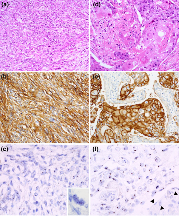
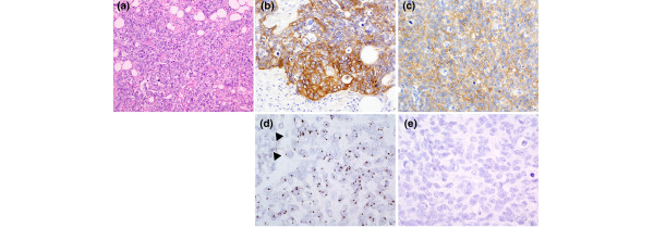
Method: Twenty-five metaplastic breast carcinomas were immunohistochemically analyzed using a monoclonal antibody (31G7) for EGFR and two antibodies for HER2 (Herceptest and CB11) and scored using the Herceptest scoring system. Gene amplification was evaluated by chromogenic in situ hybridization using Zymed Spot-Light EGFR and HER2 amplification probe. The results were evaluated by bright field microscopy under 40x and 63x objective lenses.
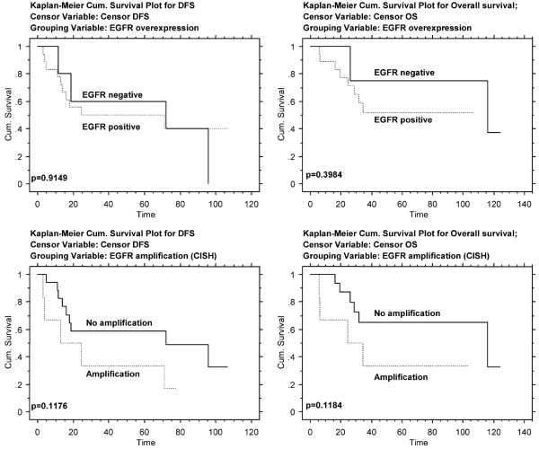
Results: Nineteen (76%) metaplastic breast carcinomas exhibited EGFR ovexpression, and among these EGFR amplification (defined either by large gene clusters or >5 signals/nucleus in >50% of neoplastic cells) was detected in seven cases (37%): three carcinomas with squamous differentiation and four spindle cell carcinomas. One case exhibited HER2 overexpression of grade 2+ (>10% of cells with weak to moderate complete membrane staining), but HER2 gene amplification was not detected.
Conclusion: Metaplastic breast carcinomas frequently overexpressed EGFR, which was associated with EGFR gene amplification in one-third of cases. Our findings suggest that some patients with metaplastic breast carcinomas might benefit from novel therapies targeting EGFR. Because most metaplastic breast carcinomas overexpress EGFR without gene amplification, further studies to evaluate EGFR activating mutations are warranted.
Figures
Similar articles
-
Molecular analysis of metaplastic breast carcinoma: high EGFR copy number via aneusomy.Mol Cancer Ther. 2008 Apr;7(4):944-51. doi: 10.1158/1535-7163.MCT-07-0570. Mol Cancer Ther. 2008. PMID: 18413808 Free PMC article.
-
EGFR amplification and lack of activating mutations in metaplastic breast carcinomas.J Pathol. 2006 Aug;209(4):445-53. doi: 10.1002/path.2004. J Pathol. 2006. PMID: 16739104
-
EGFR gene amplification in breast cancer: correlation with epidermal growth factor receptor mRNA and protein expression and HER-2 status and absence of EGFR-activating mutations.Mod Pathol. 2005 Aug;18(8):1027-33. doi: 10.1038/modpathol.3800438. Mod Pathol. 2005. PMID: 15920544
-
Epidermal growth factor family of receptors in preneoplasia and lung cancer: perspectives for targeted therapies.Lung Cancer. 2003 Aug;41 Suppl 1:S29-42. doi: 10.1016/s0169-5002(03)00137-5. Lung Cancer. 2003. PMID: 12867060 Review.
-
Emerging technologies for assessing HER2 amplification.Am J Clin Pathol. 2009 Oct;132(4):539-48. doi: 10.1309/AJCPV2I0HGPMGBSQ. Am J Clin Pathol. 2009. PMID: 19762531 Review.
Cited by
-
Demystifying basal-like breast carcinomas.J Clin Pathol. 2007 Dec;60(12):1328-32. doi: 10.1136/jcp.2006.041731. Epub 2007 May 11. J Clin Pathol. 2007. PMID: 17496191 Free PMC article. Review.
-
Metaplastic Carcinoma of the Breast Is More Aggressive Than Triple-negative Breast Cancer: A Study From a Single Institution and Review of Literature.Clin Breast Cancer. 2017 Aug;17(5):382-391. doi: 10.1016/j.clbc.2017.04.009. Epub 2017 Apr 26. Clin Breast Cancer. 2017. PMID: 28529029 Free PMC article. Review.
-
Molecular analysis of metaplastic breast carcinoma: high EGFR copy number via aneusomy.Mol Cancer Ther. 2008 Apr;7(4):944-51. doi: 10.1158/1535-7163.MCT-07-0570. Mol Cancer Ther. 2008. PMID: 18413808 Free PMC article.
-
ctDNA dynamics: a novel indicator to track resistance in metastatic breast cancer treated with anti-HER2 therapy.Oncotarget. 2016 Oct 4;7(40):66020-66031. doi: 10.18632/oncotarget.11791. Oncotarget. 2016. PMID: 27602761 Free PMC article.
-
Single-molecule imaging and fluorescence lifetime imaging microscopy show different structures for high- and low-affinity epidermal growth factor receptors in A431 cells.Biophys J. 2008 Feb 1;94(3):803-19. doi: 10.1529/biophysj.107.112623. Epub 2007 Sep 21. Biophys J. 2008. PMID: 17890389 Free PMC article.
References
-
- Suo Z, Yang H, Mei Q, Skovlund E, Cui J, Nesland JM. Type 1 protein tyrosine kinases in Chinese breast carcinomas: a clinicopathologic study. Int J Surg Pathol. 2001;9:177–187. - PubMed
Publication types
MeSH terms
Substances
Grants and funding
LinkOut - more resources
Full Text Sources
Medical
Research Materials
Miscellaneous
