

Quantifying bedside-derived imaging of microcirculatory abnormalities in septic patients: a prospective validation study
- PMID: 16280059
- PMCID: PMC1414044
- DOI: 10.1186/cc3809
Quantifying bedside-derived imaging of microcirculatory abnormalities in septic patients: a prospective validation study
Abstract
Introduction: The introduction of orthogonal polarization spectral (OPS) imaging in clinical research has elucidated new perspectives on the role of microcirculatory flow abnormalities in the pathogenesis of sepsis. Essential to the process of understanding and reproducing these abnormalities is the method of quantification of flow scores.
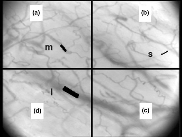
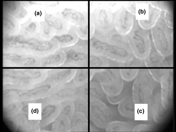
Methods: In a consensus meeting with collaboraters from six research centres in different fields of experience with microcirculatory OPS imaging, premeditated qualifications for a simple, translucent and reproducible way of flow scoring were defined. Consecutively, a single-centre prospective observational validation study was performed in a group of 12 patients with an abdominal sepsis and a new stoma. Flow images of the microcirculation in vascular beds of the sublingual and stoma region were obtained, processed and analysed in a standardised way. We validated intra-observer and inter-observer reproducibility with kappa cross-tables for both types of microvascular beds.
Results: Agreement and kappa coefficients were >85% and >0.75, respectively, for interrater and intrarater variability in quantification of flow abnormalities during sepsis, in different subsets of microvascular architecture.
Conclusion: Semi-quantitative analysis of microcirculatory flow, as described, provides a reproducible and transparent tool in clinical research to monitor and evaluate the microcirculation during sepsis.
Figures
References
-
- Sakr Y, Dubois MJ, De Backer D, Creteur J, Vincent JL. Persistent microcirculatory alterations are associated with organ failure and death in patients with septic shock. Crit Care Med. 2004;32:1825–1831. - PubMed
-
- Ince C. Microcirculation in distress: a new resuscitation end point. Crit Care Med. 2004;32:1963–1964. - PubMed
-
- Slaaf DW, Tangelder GJ, Reneman RS. A versatile incident illuminator for intravital microscopy. Int J Microcirc Clin Exp. 1987;6:391–397. - PubMed
-
- Mathura KR, Alić L, Ince C. Initial clinical experience with OPS imaging for observation of the human microcirculation. In: Vincent JL, editor. Yearbook of Intensive Care and Emergency Medicine. New York: Springer-Verlag; 2001. pp. 233–245.
-
- Mathura KR, Bouma GJ, Ince C. Abnormal microcirculation in brain tumours during surgery. Lancet. 2001;358:1698–1699. - PubMed
Publication types
MeSH terms
LinkOut - more resources
Full Text Sources
Other Literature Sources
Medical
